archive page

The Complete Guide to Tongue Ties and Frenotomy: What Every Parent Should Know

Welcoming a baby into the world is an incredible experience. But finding out your sweet new baby has a tongue tie can quickly send any parent into a whirlwind of stress and anxiety.

Fortunately, in most cases, a doctor can fix a child’s tongue tie quickly and easily with a minor procedure called a frenotomy

If you’re considering a frenotomy for your child’s tongue tie, here’s what you need to know.

What Is a Tongue Tie and When Does It Require Treatment?

A tongue tie (also called ankyloglossia) involves a small piece of tissue called the lingual frenulum, which connects the underside of the tongue to the bottom of the mouth. A normal frenulum allows the tongue to move fairly freely, but sometimes people are born with their frenulum in a position that hinders the tongue’s range of motion, “tying” the tongue to the floor of the mouth.

To be clear, when a person has a tongue tie, there’s nothing wrong with the tongue itself. It’s simply anchored too tightly, and that anchor won’t let the tongue move up or forward like it should. You can think of it like a rope holding the tongue down. As soon as that rope is cut, the tongue is free to function normally.

Parents often notice a tongue tie when their child has trouble breastfeeding or learning to speak clearly. A doctor can diagnose a tongue tie with a physical exam, but depending on the severity, it may or may not require surgical treatment.

If you know or suspect your child has a tongue tie, you may wonder whether they need surgical intervention. Generally speaking, here’s the rule of thumb we follow:

  • If a person can extend their tongue about five millimeters past their teeth and the condition doesn’t cause any issues with function, we don’t recommend surgery.
  • If the tongue tie is more severe and interferes with necessary oral functions, we recommend correcting it surgically with a frenotomy.

What Is a Frenotomy?

A frenotomy is a simple surgical cut that releases the tongue, allowing it to move freely.

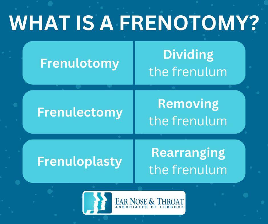
It’s also sometimes called a frenulotomy, frenulectomy, or frenuloplasty, all Latin terms that refer to very similar things:

  • Frenulotomy means dividing the frenulum.
  • Frenulectomy means removing the frenulum.
  • Frenuloplasty means rearranging the frenulum.

During a frenotomy, we use sterile scissors to gently snip the frenulum under the tongue. Since the tissue has minimal nerves and blood flow, it barely bleeds, and the patient feels minimal discomfort.

Although it might be difficult to imagine your baby or young child undergoing any surgical procedure, it really is quick and simple. In fact, it’s a lot like getting their ears pierced! It’s over within seconds and doesn’t require anesthesia — just holding their head still for a few moments.

Afterward, you can immediately nurse your child to help calm and comfort them. Older children can have a drink right away.

Infographic: The Complete Guide to Tongue Ties and Frenotomy: What Every Parent Should Know

Can a Doctor Complete a Frenotomy In-Office?

We typically perform frenotomies in the office. Newborns and infants tolerate a frenotomy very well, and we can easily hold them still for the few moments it takes to safely snip the frenulum. 

A five-year-old may not be as cooperative. If a child isn’t able to sit still for the procedure, it could be dangerous for us to attempt it. In these cases, we can administer lidocaine to numb the area and help calm the child, ensuring their safety.

If a patient is already scheduled for another surgical procedure, like a tympanoplasty (ear tube placement) or tonsillectomy (tonsil removal), we can complete the frenotomy at the same time in the operating room while they’re under anesthesia.

What About an Upper Lip Frenulectomy?

The lingual frenulum isn’t the only frenulum of the body. Another frenulum sits where the center of your upper lip meets your gum. (If you lift your tongue above your front two teeth, you can feel the small, stretchy band of tissue there.)

When this tissue attaches to the upper lip in a way that limits movement or interferes with the teeth, it’s called an upper lip tie.

Unlike a tongue tie, an upper lip tie doesn’t usually cause problems with oral functions like swallowing or speaking. Since a labial (lip) frenulectomy — the surgery to fix an upper lip tie — is more complex than a lingual (tongue) frenotomy, involving more bleeding and a less certain outcome, treatment isn’t necessary or helpful in the absence of symptoms.

In some cases, however, a very large upper lip tie can interfere with nursing or cause unwanted spacing or cosmetic issues between the upper front teeth.

Because properly correcting an upper lip tie involves raising a mucosal flap and removing some tissue between the teeth, we recommend seeing an experienced oral surgeon or pediatric dentist for assessment and treatment. They have specific medical expertise in these oral structures and offer the best path toward a successful outcome.

Early Intervention Is Key 

A tongue tie often comes to light when a lactation consultant or speech therapist identifies it as the source of a child’s nursing or speech problems. If you or your child’s specialist notice a possible tongue, we recommend consulting your pediatrician and/or ENT as early as possible. Tongue ties can interfere with feeding and impact a child’s oral functions as they grow and develop, so it’s a good idea to get them assessed and treated promptly.

When all is said and done, a frenotomy is a simple and effective solution to tongue ties, and nothing to be worried about. It’s quick, it’s easy, and it gives your child perfectly normal tongue function without any long-term consequences.

Parathyroid Exploration: From Surgery Prep to Recovery 

Symptoms like weak bones, kidney stones, joint pain, and mental fog can make it very difficult to thrive in daily life. Few people realize an overactive parathyroid gland may be the culprit.

If you have one or more overactive parathyroid glands, you’ll likely need one of two types of surgery: parathyroid exploration or minimally invasive parathyroidectomy (MIP). Keep reading to find out if it may be necessary for you and what you can expect from the procedure and its outcome.

Why Do People Need Parathyroid Surgery?

First, let’s start with the basics:

Your thyroid is a butterfly-shaped gland located at the front of your neck. Behind it are four small glands called parathyroid glands. There are two on each side: an inferior (lower) and a superior (higher) gland.

Your parathyroid glands are about two to four millimeters in diameter, smaller than a grain of rice. Though tiny, they play a significant role in your bodily functions. These tiny glands make a hormone called parathyroid, which regulates the calcium levels in your bones and blood.

Sometimes, one or more of these glands (usually just one) can balloon to 20 or 30 times its normal size. That’s like growing from the size of a grain of rice to a marble, or larger! 

The enlarged gland — called a parathyroid adenoma — goes completely rogue, producing a nonstop supply of parathyroid hormone. The result is a condition called hyperparathyroidism, which is the most common reason we perform MIP or parathyroid exploration surgery. 

Infographic: Parathyroid Exploration: From Surgery Prep to Recovery

What Are Common Hyperparathyroidism Symptoms?

When you have hyperparathyroidism, all the excess parathyroid hormone in your body strips the calcium from your bones, which you then excrete in your urine. This can lead to a host of unpleasant symptoms, including:

  • Osteoporosis
  • Kidney stones
  • Organ damage
  • Depression
  • Brain fog
  • Other mental health issues

Hyperparathyroidism has earned quite a memorable nickname as “the illness of bones, stones, and abdominal groans.” Some people even add “psychic moans” to the list, since the excess hormone can affect a person’s mental health.

How Is Hyperparathyroidism Diagnosed?

To diagnose hyperparathyroidism, we conduct biochemical testing, which gives us more insight into your calcium, parathyroid hormone, and vitamin D levels. 

If these blood tests indicate hyperparathyroidism is present, we then use localization studies to find which parathyroid gland is enlarged. This process sometimes includes a thyroid ultrasound, a radioactive tracer study called a sestamibi (MIBI) scan, or a 4D CT scan.

Thankfully, our ability to find the enlarged gland has improved tremendously over the past several years, reducing the need for locating the offending gland during a parathyroid exploration.

MIP vs. Parathyroid Exploration

Whenever possible, we prefer to use minimally invasive parathyroid surgery (MIP) to treat hyperparathyroidism. This procedure is much more common now because of the improvements in our ability to pinpoint the location of the problem-causing gland.

MIP is a quick surgical procedure that allows us to use very small incisions and remove the enlarged gland with precision, all with minimal risk to you. 

Sometimes, however, we’re not able to locate the problematic parathyroid gland before surgery. In these cases, we may need to perform a parathyroid exploration surgery to look at all four glands and determine where the problem lies.

What to Expect Before the Procedure

Before parathyroid surgery, you’ll need biochemical testing to confirm hyperparathyroidism. If the results are positive, you’ll undergo localization studies like those mentioned above to pinpoint the gland(s) we need to remove.

What to Expect During the Procedure

Whether we use MIP or parathyroid exploration surgery, it’s usually an outpatient procedure. Depending on how many glands we need to look at, the surgery takes about 20 minutes to 1.5 hours. It’s always best to choose a high-volume surgeon; their familiarity with the procedure not only reduces the likelihood of complications but can also mean a quicker surgery.

For parathyroid exploration, we use a thyroidectomy incision in the front center of the neck to access your parathyroid glands. If we’re able to use MIP, then we can make much smaller incisions to remove the overactive gland.

After we remove the gland, we use invisible sutures to close the skin and apply Steri-Strips to securely bring both sides of the incision together and seal the opening. A wound drain is not necessary for this procedure.

What to Expect After the Procedure

Once we remove the enlarged gland, your calcium and parathyroid hormone levels should return to normal within as little as 20 minutes.

Whether we need look at all four glands with a parathyroid exploration or we’re able to do the minimally invasive parathyroid surgery, post-op recovery is minimal.

After a period of observation, most patients will be able to go home the day of their procedure. We ask you to check into the office the next day for a follow-up to make sure there are no complications. The most common complaints after parathyroid surgery are soreness at the incision site and slight neck pain, which is the result of your head positioning during surgery. Both of these complaints usually only last a day or two; then, you are back to normal activity.

Tympanoplasty vs. Myringoplasty vs. TM Patch: Differences Explained

In many cases, a perforated eardrum will heal on its own. But if yours doesn’t, you may need surgery to repair the damage to protect your middle ear and improve your hearing.

A tympanoplasty, myringoplasty, and TM patch (also called a paper patch) are all effective methods for repairing a hole in the eardrum. Which one is best for you depends on the size of the hole, your age, and your doctor’s recommendations.

To help you move forward with your treatment, we’ll compare a paper patch vs. a myringoplasty vs. a tympanoplasty so you can fully understand each procedure.

Why Is It Important to Treat Tympanic Membrane Perforations?

A tympanic membrane perforation is a medical term for a hole in the eardrum. These can result from severe or repeated ear infections or an injury, such as getting hit in the ear or pushing an object into your ear, like a Q-tip.

Your eardrum separates your outer ear from your middle ear, and it’s very important to maintain this divide.

As soundwaves hit your eardrum, the membrane sends vibrations through the little bones of your middle ear on to your inner ear and brain, where they’re interpreted as sound. A hole in your eardrum, especially a large hole, interferes with these vibrations.

Additionally, the eardrum protects your middle ear from water, bacteria, and debris that could otherwise get in and cause infection. Repairing a lingering hole restores this important protection.

The three main procedures an ENT may use to repair a hole in your eardrum are a tympanoplasty, myringoplasty, and a TM patch.

What Is a Tympanoplasty?

A tympanoplasty is a surgery to repair a large hole in your eardrum, typically caused by chronic infections or trauma.

During the operation, we enter through your ear canal or make a small incision behind your ear to lift the eardrum and access your middle ear. Once inside, we place a tissue graft underneath your eardrum to repair the hole from the inside.

A tympanoplasty is the most major of the three types of eardrum surgeries listed in this post. It takes about two to three hours to complete, and we prefer to wait until patients are at least eight years old before using it as a treatment option.

What Is a Myringoplasty?

We use a myringoplasty to repair smaller holes in the eardrum, also caused by chronic infections or trauma to the ear.

This simple operation involves repairing the surface of your eardrum instead of the inner ear. During the procedure, we go in through the ear canal and use a tiny piece of earlobe fat to plug the hole. As the hole heals over time, the fat is incorporated into the eardrum.

The entire surgery takes 10–30 minutes and is well-tolerated, even by children as young as one or two years old.

What Is a TM Patch or Paper Patch?

Like a myringoplasty, a TM patch or paper patch is a simple ear surgery that takes 10–30 minutes to complete. This procedure works best on the smallest of eardrum perforations.

During the operation, we enter through the ear canal to carefully roughen the edge of the eardrum perforation. We then place a small paper patch or Medtronic EpiDisc containing growth factors over the hole to enhance the healing process.

Infographic: Tympanoplasty vs. Myringoplasty vs. TM Patch: Differences Explained

Paper Patch vs. Myringoplasty: What’s the Difference?

When comparing a paper patch vs. a myringoplasty, it’s easy to get lost in the technical details, and it’s not always clear what the difference is. Although they are similar procedures, there’s one main difference:

  • A paper patch is for the smallest perforations and repairs the eardrum’s surface by placing a tiny patch over the hole to help it heal. 
  • A myringoplasty is for small to moderate holes and involves inserting a fat graft through the hole, so it’s both in the middle ear and on the eardrum’s surface.

How to Decide Which Procedure Is Right for You

It’s always best to see an ENT for a personalized recommendation, but here’s the bottom line:

  • If the perforation is small, we’ll most likely recommend a myringoplasty or paper patch, especially if the patient is a child. Both procedures will encourage the hole to heal quickly and can be done when a child is young instead of waiting until they’re old enough to have more major ear surgery.
  • If the perforation is large, a myringoplasty may still work to repair it. If that’s the case, we’ll recommend a myringoplasty over a tympanoplasty so you don’t have to endure a major surgery. A paper patch is exclusively for smaller perforations, so it’s not an option in this case.

Even a small hole in your eardrum can enlarge and cause more issues down the road. We prefer to catch the problem early so we can repair it with a simple myringoplasty or TM patch and avoid the need for more major middle ear surgery in the future.

All three ear surgeries mentioned above are safe ways to repair a perforated eardrum, but we may recommend one over the others depending on your situation.

Vocal Cord Stripping: Before, During, and After Your Procedure 

If you’ve dealt with vocal cord conditions like irreversible swelling, polyps, or non-invasive cancer, you’re no stranger to the symptoms: chronic sore throat, difficulty speaking, or the irritating sensation of something stuck in your throat.

In certain cases, vocal cord stripping can be a safe and effective solution. 

Let’s take a closer look at this procedure to find out what it is and how it can help.

What Is Vocal Cord Stripping and What Are the Benefits?

Your vocal cords are made up of two ligaments on either side of your larynx (or voice box), covered by muscle wrapped in a delicate, fine layer of mucosal tissue. This outer tissue can sometimes become damaged, inflamed, or diseased, necessitating vocal cord stripping.

Vocal cord stripping is a surgery that involves gently removing the outermost layer of vocal cord tissue when it has become irreversibly diseased. The process removes the damaged cells and allows new tissue to grow back healthier in its place.

We might recommend vocal cord stripping if you have specific vocal cord conditions, including:

  • Irreversible swelling
  • Polyps
  • Dysplasia (abnormal, pre-cancerous cells)
  • Carcinoma in situ (non-invasive cancer that hasn’t spread to the nearby tissues)

For instance, if you have dysplasia, which is abnormal, pre-cancerous cells on the vocal cord, vocal cord stripping removes those cells before they progress to cancer.

Infographic: Vocal Cord Stripping: Before, During, and After Your Procedure

What Happens Before the Procedure?

Vocal cord stripping isn’t the first line of treatment. Depending on the ailment, we’ll recommend other, more conservative medical treatments for vocal cord issues first, such as:

  • Antibiotics
  • Antifungal medications
  • Oral steroids
  • Reflux medications
  • Behavioral modifications like avoiding cigarettes and alcohol

If none of these treatments work and your vocal cords reach the point of irreversible damage, vocal cord stripping will likely be our next step.

What Happens During Vocal Cord Stripping?

Don’t worry — this procedure takes place under general anesthesia, so you won’t feel a thing!

Once the anesthesia takes effect, we insert a scope into your mouth to see your voice box and vocal cords under a microscope.

We then use long, tiny forceps to grasp the vocal cord and a pair of tiny micro-laryngeal scissors to strip off the damaged outer layer of delicate tissue containing the abnormal cells, taking care not to injure the deeper parts of the vocal cord.

The entire surgery takes about 15–30 minutes.

What Happens After Vocal Cord Stripping?

After vocal cord stripping, you’ll need complete vocal rest — or in other words, no talking — for three to seven days. Beyond that, you’ll also want to avoid abusing your voice with shouting or excessive talking for several months, since these activities strain your vocal cords. 

Depending on the patient, we may recommend the following post-op treatments to help with healing and prevent future injury or disease:

  • Speech therapy may help you develop practical vocal techniques to gradually recover your full voice function and avoid injuring your vocal cords in the future.
  • Anti-reflux medications like Nexium can help prevent further injury to your vocal cords from stomach acid. 
  • Behavioral changes like stopping smoking or drinking will help your throat heal faster and help prevent the need for treatment again in the future.

Talk to Your Doctor to Find Out if Vocal Cord Stripping Is Right for You

If the delicate lining of your vocal cords becomes irreversibly damaged or diseased, it’s important to treat it for your comfort and health. If medications and behavioral changes fail, vocal cord stripping may be the best solution. 

Always speak with your ENT about any vocal cord issues you’re having so you can find out the best next step for your health!

Before, During and After Your In-office Turbinate Resection

Hearing the words “turbinate resection” come out of your doctor’s mouth might make you want to run. We get it. It sounds intimidating!

Fortunately, a turbinate resection isn’t nearly as scary as you think.

In this post, find out how simple and easy a turbinate resection really is and how it can help you start breathing more comfortably.

Why Turbinates Matter

Let’s quickly review what turbinates are and why they’re important.

Turbinates are small, spongy, finger-like structures that lie along the length of your nasal wall. Each of your nasal passages holds three turbinates: the superior, middle, and inferior turbinates. Their job is to warm and humidify the air you inhale through your nose and regulate nasal breathing.

The trouble comes when allergies or infections make your turbinates swell. This swelling, especially of the inferior turbinate, constricts the nasal airway and makes it difficult to breathe through your nose. If the swelling becomes frequent, you can end up with a chronic breathing dilemma on your hands.

Common Misconceptions About the Turbinate Resection Procedure

Because a turbinate resection sounds intimidating, some misconceptions have grown up around it. Here are some of the most common:

  • You need general anesthesia. Although some ENTs may use general anesthesia for a turbinate resection, we simply employ local anesthesia for our patients. They don’t experience any discomfort during the procedure, which is simple, easy, and very well-tolerated.
  • It’s major surgery. On the contrary, a turbinate resection is a minimally-invasive procedure we complete quickly and easily at our office. Sometimes we even do it in conjunction with other procedures, like a balloon sinuplasty or VivAer
  • Recovery takes several days. Recovery from a turbinate resection is very quick and easy. You can resume work and regular activities the day after your procedure. 
Infographic: Before, During, and After Your In-Office Turbinate Resection

What Is a Turbinate Resection?

If swollen turbinates continually restrict your breathing, we may recommend a turbinate resection to alleviate the problem. 

Years ago, completely or partially cutting off the inferior turbinate was an accepted practice. However, that’s no longer recommended, and it’s not what we do in our office.

Today, a turbinate resection is a much more conservative procedure, simply reducing the inferior turbinate’s size by a few millimeters. Although it’s a minimal change, it can tremendously impact your nasal breathing and help with issues like sinus infections and snoring.

What to Expect During a Turbinate Resection

During a turbinate resection, you’ll be awake but won’t feel any pain.

First, we’ll gently make a small incision at the front of the turbinate. 

Then, we’ll insert a tiny, two-millimeter blade into the incision and remove some of the turbinate’s interior tissue to reduce its size. The process is similar to liposuction, just on a much smaller level.

By removing a small portion of the spongy tissue, the turbinate can’t swell as much and block your nasal passages, allowing you to breathe easier.

What Is the Goal of a Turbinate Resection?

If you get sinus infections or have allergy symptoms, you may already be familiar with swollen turbinates, blocked nasal passages, and difficulty breathing.

The ultimate goal of a turbinate resection is to reduce the amount your inferior turbinates can swell. Then, those pesky allergies and sinus infections can’t interfere with your breathing so much. You may even notice reduced snoring!

Turbinate Resection: Final Thoughts

If your ENT has recommended a turbinate resection, you might feel a little nervous. This is normal, but we hope the above description of the simplicity of the procedure will set your mind at ease. You deserve to breathe freely!

Heterogeneous Thyroid: Symptoms, Causes, and Treatment

Suppose you’ve been having trouble swallowing, or you feel pressure on your neck when lying flat. Or maybe you have a visible lump on one side of your neck, and you’re not quite sure what it is.

If any of those scenarios apply to you, run, don’t walk, to consult a doctor. They could indicate the presence of a heterogeneous thyroid.

What Is a Heterogeneous Thyroid?

This is often a difficult term for patients to understand, but very simply, a heterogeneous thyroid is an irregular type of thyroid gland. It’s brought on by autoimmune thyroiditis, in which your body’s immune system can’t differentiate between your own cells and foreign cells. In these cases, your body creates antibodies that attack the healthy tissues in your thyroid glands, much like Hashimoto’s or Grave’s disease.

A heterogeneous thyroid becomes overactive (hyperthyroidism) or underactive (hypothyroidism). In either case, the thyroid produces irregular amounts of the hormone thyroxine, which affects almost all your body’s systems, including digestive functions, metabolism, bone health, and muscle control.

As a result of this condition, you might experience symptoms like hyperactivity or extreme fatigue. Doctors can prescribe medications to regulate your hormone levels and help treat these symptoms. Complete thyroid gland removal may also help you feel better. Often, with complete gland removal, any pressure in the neck also subsides.

Infographic: Heterogeneous Thyroid: Symptoms, Causes & Treatment

What Causes a Heterogeneous Thyroid?

A heterogeneous thyroid is an autoimmune disease — a condition in which your immune system misfires and attacks your own body. Unfortunately, there’s nothing you can do to prevent a heterogeneous thyroid. Doctors don’t know exactly what causes autoimmune diseases, but women are more likely to get them.

Treatment Options for a Heterogeneous Thyroid

If you have symptoms of a heterogeneous thyroid, the first step is to be evaluated by an ENT doctor or an endocrinologist, an expert in the study of your body’s hormones.

The doctor will check your thyroid levels and perform a thyroid ultrasound. They may also perform a test like a nuclear medicine scan to check for abnormal tissue growths like thyroid nodules or tumors.

Whether the doctor finds that your thyroid is heterogeneous or homogeneous (healthy), they’ll pay special attention to any internal nodules. If they have concerns about possible thyroid cancer, they may want to perform a needle biopsy to extract a tissue sample for further testing.

Depending on their findings, they may also recommend removing the entire thyroid gland; a surgical procedure called a thyroidectomy.

What Should I Do Next?

Your doctor will recommend an individual treatment plan based on the results of their initial evaluation. They may prescribe medications to balance your hormones if you have an underactive or overactive thyroid. However, if the medicine isn’t effective, a thyroidectomy may be preferred.

Similarly, several other factors could prompt your doctor to remove one of your thyroid glands, including its overall size, the presence of nodules, or the presence of Hashimoto’s disease or Grave’s disease.

Regardless, the best thing you can do is seek expert treatment recommendations from a medical professional. They will help you determine the proper next steps based on your symptoms and diagnosis.

An ENT Doctor Will Ensure Safe and Effective Treatment for a Heterogeneous Thyroid

Understandably, if you’re experiencing symptoms of a heterogeneous thyroid, you might be anxious or worried about a diagnosis. Fortunately, you’re in good hands with an ENT doctor. They can guide you through the proper steps to address your symptoms and ensure you get the treatment you need to feel your best again.

The Best Sleeping Position for Sleep Apnea Sufferers

As you age, your likelihood of being diagnosed with sleep apnea increases — people over 65 are twice more likely to need CPAP machines than younger people because of the loss of muscle tone that comes with aging. While this may be frustrating, rest assured that it’s just another part of the natural aging process.

What also may seem out of your control is your sleeping position, which could be leading to sleep apnea’s more harmful side effects. This is a topic we’ve been asked about often over the years — believe it or not, some patients have even sewed tennis balls to the back of their pajamas to keep from sleeping on their back!

The fact is, we only have so much control over our sleeping position, but there are minor things we can do to alter it for our health’s sake. Let’s look at how your sleeping position could be causing side effects like snoring and abnormal breathing and identify the best sleeping position for sleep apnea.

What Is Sleep Apnea?

Sleep apnea is a sleep disorder in which your breathing repeatedly stops and starts throughout the night. The most common type of sleep apnea is called obstructive sleep apnea (OSA), which occurs when the throat muscles relax.

When you sleep, your throat and tongue muscles relax. If you sleep on your back, gravity makes your tongue collapse into your airway, restricting airflow and causing that classic snoring noise we’re all familiar with.

Thankfully, sleep apnea treatments like Inspire are highly effective. This tiny implantable device stimulates the hypoglossal nerve, which controls tongue movement and other airway muscles, keeping your airway open while you sleep.

Other small lifestyle changes, like your sleep position, may also negate some of the adverse side effects of sleep apnea — thus why I’ve heard the question so many times throughout my career.

How Does Sleeping on Your Side Affect Sleep Apnea?

If you suffer from sleep apnea, one of the best ways you can manage it is by sleeping on your side. Whether you sleep on your right or left, your tongue won’t fall back very much, and your airway will remain more open, enabling you to breathe correctly. A recent study found that people with sleep apnea who sleep on their side are less likely to experience disordered breathing at night.

Although there are several different ways you can sleep on your side, the fetal position — sleeping on your left or right side with your legs curled inward toward your body — is the most popular. Research indicates that sleeping in the fetal position may reduce snoring and airway blockage among people with sleep apnea.

How Does Sleeping on Your Stomach Affect Sleep Apnea?

Similarly, when you sleep on your front or belly, your tongue won’t fall back into your throat as much as it would if you were on your back. According to the Better Sleep Council, only about 17% of people sleep on their stomachs, so while it may not be the most desirable sleeping position for most people, it will keep your airway clear at night and help prevent the snoring associated with sleep apnea.

How Does Sleeping on Your Back Affect Sleep Apnea?

Sorry to break it to all you back sleepers, but sleeping on your back is the least favorable position for sleep apnea. If you’re on your back, your tongue can easily fall back into your throat and restrict your airflow. If this is your preferred sleeping position, it may take some time to adjust to a new position, but if you suffer from obstructive sleep apnea, it’s well worth the effort.

What Treatments Are Best for People With Sleep Apnea?

Infographic: The Best Sleeping Position for Sleep Apnea Sufferers

If you have sleep apnea, some great ways to reduce the negative side effects include:

  • Maintaining a healthy weight: Excess tissue in the throat can cause further obstruction and breathing issues at night. Achieving and maintaining a healthy weight will reduce the amount of tissue in your throat and may improve your breathing.
  • Avoid drinking alcohol at night: Research suggests alcohol consumption is associated with a higher risk of sleep apnea because it disrupts sleep patterns, relaxes muscles excessively, and decreases overall sleep quality. Certain prescription drugs like muscle relaxants, sedatives, and narcotics have the same effects, so try to avoid those as well.
  • Avoid sleeping on your back: Experimenting with the best sleeping positions for sleep apnea may reduce its side effects. Do your best to sleep on your side, preferably in the fetal position if that feels comfortable. If you tend to roll onto your back during the night, try placing a pillow between your legs. It may hinder your movements and keep you on your side.
  • Use evidence-based treatments: Your doctor may recommend proven treatments for sleep apnea, including a mouthguard, CPAP (continuous positive airway pressure therapy) machine, or, if you can’t tolerate CPAP, Inspire.

See an ENT Doctor for Effective Sleep Apnea Treatment

If you’re struggling with disrupted breathing and snoring caused by sleep apnea, switching to an ideal sleeping position for sleep apnea, like side or stomach sleeping, may help. If not, a trusted ENT doctor can help you identify other solutions.

A Surgeon Explains Deviated Septums & Sleep Apnea

A deviated septum and sleep apnea are two common medical conditions that affect your ability to breathe correctly. But are they related? Can a deviated septum cause sleep apnea?

The short answer is no, a deviated septum can’t cause sleep apnea. The long answer is still no, but it includes some important details about how a deviated septum can affect sleep apnea sufferers, even though it doesn’t cause their condition.

What Is a Deviated Septum?

The septum is the central wall in your nose that separates the nasal cavity into two sections: right and left. The front portion is made of cartilage, and the back is made of bone.

The septum should run straight down the center of your nose, but sometimes the bone in the back pushes the cartilage into an uneven position. This is what medical professionals call a deviated septum.

A graphic showing a normal nasal septum vs a deviated septum to explain the relation of deviated septums and sleep apnea.

The deviation doesn’t always occur on just one side, either. Sometimes it affects both sides, with the septum protruding in one direction in the front and the opposite direction in the back.

Symptoms of a deviated septum include:

If your nose is visibly crooked and/or you’re experiencing some of the above symptoms, a deviated septum could be the cause. An ENT doctor can check for a deviated septum with a quick and painless exam, which includes visually inspecting your septum with a small light.

Infographic: A Surgeon Explains Deviated Septums & Sleep Apnea

What Causes a Deviated Septum?

A deviated septum happens for various reasons, including the following:

  • A congenital condition: It’s not uncommon for people to have a deviated septum from birth. It may result from a difficult birthing process or just a slight malformation of the nasal structure.
  • Gradual growth and development: As your nose grows, your septum does too. Sometimes, it can grow unevenly to one side, perhaps if the underlying bone is slightly crooked. As the septum grows, the deviation becomes more pronounced.
  • A nasal injury: Any trauma to the nose, like a sports injury, car accident, or a fall, can cause the septum to deviate.

What Is Sleep Apnea?

Sleep apnea is a medical condition where breathing stops and starts many times while you sleep. The most common type of sleep apnea is obstructive sleep apnea, in which your upper airway becomes blocked by throat collapse or by your relaxed tongue or tonsils falling backward into your airway.

After you stop breathing, you startle awake and your muscles regain their tone, opening your airway. Then you quickly fall back to sleep. Typically, people with sleep apnea don’t even realize they’re waking up dozens to hundreds of times throughout the night. They just feel exhausted in the morning.

All of this pseudo-waking is your body’s way of keeping you alive. However, sleep apnea still has some seriously harmful health effects

  • It prevents you from getting quality sleep: Constantly being startled awake at night prevents you from falling into a deep sleep or rapid eye movement (REM) sleep. As a result, you never truly get a full night’s rest, and you feel tired all the time.
  • It decreases your body’s oxygen levels: Reduced oxygen levels strain your heart, lungs, and brain, ultimately shortening your lifespan.
  • It reduces your overall quality of life: Not getting enough sleep is linked to health problems like obesity, diabetes, cardiovascular disease, and decreased immune function.

To help you breathe easier and get better sleep, an ENT doctor may recommend using a CPAP (continuous positive airway pressure) machine to keep your airway open while you sleep.

How Does a Deviated Septum Affect People Who Have Sleep Apnea?

So, how are a deviated septum and sleep apnea related?

People can have a deviated septum and sleep apnea simultaneously. And while a deviated septum doesn’t cause sleep apnea, it can exacerbate sleep apnea symptoms and make treatment more difficult.

A crooked septum causes turbulent airflow through the nose, potentially worsening throat collapse. Difficulty breathing through the nose also tends to increase mouth breathing, which can increase snoring and worsen sleep quality (for both you and your partner!). A deviated septum also makes you more susceptible to nasal congestion and sinus infections, which can worsen sleep quality and sleep apnea symptoms. 

Will Fixing a Deviated Septum Improve Sleep Apnea?

If your ENT diagnoses you with a deviated septum, they might recommend a septoplasty, which is minor surgery to straighten the septum.

Straightening the septum will improve nasal airflow, but it won’t entirely cure sleep apnea. However, it will improve your nasal breathing during the day and potentially reduce your snoring and the turbulent airflow worsening your throat collapse at night.

It can also help you tolerate a CPAP machine better while you sleep, which makes an enormous difference in your sleep apnea symptoms.

What to Do if You Suspect a Deviated Septum or Sleep Apnea

While a deviated septum and sleep apnea are two separate medical conditions, they both cause disordered breathing — which no one wants.

If you suspect you have undiagnosed sleep apnea, it’s important to see a doctor. A simple sleep test will make things clear. Undiagnosed sleep apnea is dangerous, but a CPAP machine or other treatment option can change (and save) your life.

If you think you have a deviated septum, it’s also a good idea to see an ENT doctor, especially if you’re having trouble tolerating your CPAP machine. Suffering with a deviated septum is uncomfortable and can make symptoms of sleep apnea worse, but a diagnosis is quick and easy.

As we mentioned, straightening a deviated septum with a septoplasty won’t cure sleep apnea. But it will improve your breathing during the day, boost breathing during exercise, and potentially reduce disordered breathing at night.

Sudden Hearing Loss in One Ear: Causes and Treatments

If you have sudden hearing loss in one ear, you’re right to be concerned, but it’s best not to jump to conclusions.

Sudden hearing loss in one ear is relatively uncommon, but in most instances, it’s not permanent and is treatable. Here’s what you need to know to achieve the best chances of restoring your hearing.

How Serious Is Sudden Hearing Loss in One Ear?

Sudden hearing loss can happen to anyone, but it’s almost always in one ear, not both. 

If you experience sudden hearing loss, you might notice it when you first wake up in the morning, or increasingly over two or three days. It might become apparent when you try to use the phone or your earbuds. Some people may even hear a popping sound before they lose hearing in one ear, while others experience vertigo or hear ringing in their ears.

Many people who experience sudden hearing loss in one ear worry that they might lose hearing in their other ear, too. Fortunately, people don’t commonly lose hearing in their other ear, so that’s not something you have to worry about.

However, sudden hearing loss in one ear is still considered a medical emergency, and you should see a doctor immediately. 

An ENT doctor (or audiologist) can test your hearing with an exam called pure tone audiometry, and they can evaluate whether your hearing loss is due to an obstruction like ear wax or fluid buildup or nerve damage in the inner ear. They may perform an MRI to rule out a tumor and other neurological problems as the cause.

According to the Hearing Loss Association of America, about half of all people with sudden hearing loss will recover some or all of their hearing spontaneously within a week or two of losing it. The odds improve if you see a doctor. About 85% of those who receive treatment from an ENT recover at least some of their hearing.

What Are Some Common Causes of Sudden Hearing Loss in One Ear?

Infographic: Sudden Hearing Loss in One Ear: Causes and Treatments

It’s not easy to determine what causes sudden hearing loss, and doctors can typically only pinpoint a specific cause about 15% of the time. In most cases, the hearing loss is caused by some type of harm to the inner ear, such as:

  • An infection: This type of inner ear damage usually results from a viral infection, but, more rarely, it can come from a bacterial infection.
  • Vascular or blood flow problems: Disorders that cause insufficient blood flow to the ear and cochlea (the hollow, spiral-shaped bone in the inner ear) may cause symptoms like fluctuating or permanent hearing loss. Examples include heart disease or ischemia, a condition where a part of your body doesn’t get enough blood.
  • Neurological disorders: Research shows that hearing impairment can accompany a variety of neurological diseases, including multiple sclerosis, Alzheimer’s disease, Parkinson’s disease, Huntington’s disease, and autism spectrum disorder.
  • A tumor: Pressure from an acoustic neuroma, a type of tumor that grows on the ear’s balance and hearing nerve, can also cause hearing loss. This type of tumor is rare, but an ENT doctor will still want to rule it out, just in case.

Of all the possible causes of sudden hearing loss in one ear, viral infections and vascular blood flow issues are the most common. An experienced ENT can determine the cause of your hearing loss and help you identify the best type of treatment for your situation.

Treatments for Sudden Hearing Loss in One Ear

The good news is sudden hearing loss is treatable, especially if you see an ENT immediately. When you go to the doctor, they’ll consider the possible causes of your condition and help you determine the appropriate treatment based on your symptoms and medical history. 

There are usually two main treatments for sudden hearing loss in one ear:

  • Oral steroids: Oral corticosteroids are the most common treatment for hearing loss. They work by decreasing inflammation and swelling in the ear to help the nerve recover. Doctors typically prescribe them in pill form. While oral steroids may have some adverse side effects like fluid retention, upset stomach, and high blood pressure, they’re highly effective in treating hearing loss.
  • Intratympanic steroids: Sometimes, an ENT may want to deliver steroids directly into your ear. To do so, they first numb your eardrum and then use a very fine needle to inject a small amount of highly concentrated steroid medication into the middle ear. The medicine travels from the middle to the inner ear to decrease inflammation and swelling. Intratympanic steroid injections may be given in conjunction with oral steroids or as an alternative treatment for patients who can’t tolerate oral steroids.

For many years, doctors tried numerous other treatments to address sudden hearing loss, but none of them stood the test of time. The only treatment that made a significant impact was steroids. 

See an ENT Doctor Right Away

If you’re experiencing sudden hearing loss in one ear, the best course of action is to see an ENT doctor right away. While the wait-and-see approach may seem attractive, it’s important to treat sudden hearing loss as early as possible to increase your chance of recovery.

With fast and proper treatment, the likelihood of restoring your hearing is very high. Although complete hearing recovery is uncommon, most people who get treatment from an ENT right away will recover some or most of their hearing.

What Is an ENT Doctor and When Should You See One?

The human body is made up of a vast network of interconnected systems, a complex machine capable of healing itself, growing new tissue, and fighting off disease. It’s so complicated and mysterious that despite studying it for generations, we still don’t know all there is to know about it.

This is why physicians cover different fields in medicine; there’s just so much to know. Some physicians are general practitioners, establishing long-term relationships with patients and monitoring their overall health for warning signs of deterioration or disease. Other physicians specialize in one body system or area, diving deep on the subject and giving it their full attention.

An ENT is one of these specialty doctors. But what is an ENT, what do they do, and when should you see one? We’ll break it down in this post.

What Is an ENT?

ENT stands for “ear, nose, and throat” and is the general term for an otolaryngologist. Otolaryngology is the branch of medicine that diagnoses and treats disorders of the head and neck.

ENT disorders involve the ears, nose, throat, or neck, and treatments for these conditions can be both medical and surgical in nature. This is why ENTs are known by yet another name: head and neck surgeons.

ENTs are highly trained, uniquely qualified doctors who intimately understand the network of passages between your ears, nose, and throat and the conditions that affect them.

What Is an ENT’s Specialty and What Do They Treat?

Some people only think of ENTs as treating severe allergies. Which they do! But they treat many other conditions, too, some common and some not so common. ENTs treat patients of all ages, from newborns to seniors, and assist with a huge variety of issues, which we’ll look at by category below.

Ear concerns:

Nasal and sinus concerns:

Throat concerns:

Pediatric ENT issues:

Head and neck complaints:

Plastic and reconstructive surgeries:

  • Cleft lips
  • Cleft palates
  • Ear deformities
  • Reconstruction
What Is an ENT and When Should You See One Infographic

How Does ENT Care Fit in With Regular Medical Care?

Typically, you see your general practitioner at least once a year to monitor your health and pick up on any new problems. Because ENT doctors are specialists, you won’t necessarily see one unless you develop an ENT issue.

Sometimes people see an ENT because their primary care doctor notices an ear, nose, throat, or neck issue, and they know to refer the patient to an ENT for further investigation and specialized treatment. Other times, patients notice ear, nose, throat, or neck issues themselves and seek out ENT care directly.

Once you establish a relationship with an ENT, they’ll determine how often you need to come in based on your unique situation. Everyone is different, so this varies greatly from person to person and condition to condition, unlike yearly primary care appointments.

When Should You See an ENT?

As you saw above, many conditions and ailments can warrant ENT care, but you may not know whether you have one of those conditions yet. So how do you know when to visit an ENT?

It really just boils down to uncertainty. If you notice any irregularities or have any concerns regarding your head and neck area, it’s always a good idea to see an ENT specialist.

Being proactive about even minor issues can make a positive difference in your health. Plus, getting a specialist’s insight can provide the intangible but valuable benefit of peace of mind. Keep in mind, though, that certain conditions may require initial evaluation by your primary care physician, who can then give you an ENT referral.

What Is an ENT Visit Like?

What is an ENT visit like, and what should you expect? At your first appointment, you’ll provide all the typical doctor’s appointment information, like insurance, medical history, medications you’re taking, etc. In the exam room, the doctor will ask about your symptoms, and then examine the relevant area.

Our office has in-house equipment so that, if necessary, we can immediately perform additional tests, like CT scans, eardrum mobility testing, and more. We can also perform certain procedures immediately in the office if needed, such as ear tube placement.

ENT appointments usually last 30-60 minutes, though additional testing may take longer. Once your doctor reaches a diagnosis,they will discuss treatment options with you. In many cases, treatment can be completed in a single office visit. However, some conditions may require ongoing care or even surgery.

The Final Word: What Is an ENT?

So, what is an ENT? An ENT is a physician who cares for people by diagnosing and treating their ears, noses, throats, and necks. They address seemingly minor issues like tonsillitis, postnasal drip, and sinus infections, which can improve a patient’s quality of life. They also take care of more serious issues like sleep apnea and various head and neck cancers, which can increase a patient’s length of life.

ENTs play an important role in healthcare. If you’re experiencing unpleasant or concerning symptoms in your ears, nose, throat, or neck, don’t be afraid to contact an ENT office today. That’s why we’re here!

Can’t Breathe Through Your Nose When Lying Down?

Few things are as uncomfortable as being unable to breathe while you’re lying down.

If you can relate, you might assume nasal problems are causing your breathing issues. In many cases that’s true, but other medical conditions can also contribute. To help you determine what’s going on, we’ll review some of the top culprits that interfere with breathing through your nose when lying down.

Cause #1: Nasal Problems

Nasal issues are the most common reason people feel like they can’t breathe through their nose when lying down. These include:

Allergies

Allergic rhinitis, also known as seasonal allergies, can sometimes cause breathing issues at night. Here in West Texas, common allergens like ragweed and dust can wreak havoc on our lives. They make it difficult to breathe during the day, let alone at night when we lie down to rest.

Uncomfortable symptoms like nasal congestion and sneezing affect your ability to breathe. They also tend to worsen at night, especially if your sleep environment is full of allergens like pet dander or dust. As a result, you might struggle to fall asleep or stay asleep, causing even further issues like daytime drowsiness.

If treatments like over-the-counter allergy medications and saline irrigation systems aren’t helping, or if you’re experiencing new or worsening allergy symptoms, an ENT doctor can help you investigate the problem and find relief.

Nasal Polyps

Nasal polyps are soft, noncancerous growths in the nose. They aren’t painful, but they can interfere with breathing. They hang like teardrops from the lining of your nasal passages and can sometimes become inflamed, blocking your airway. When you lie down, larger growths block your nasal passages and make it hard to breathe.

Nasal polyps don’t usually develop until you’re well into adulthood. People with asthma, allergies, nasal inflammation, or repeated infections are more likely to develop them. Nasal polyps don’t always produce symptoms, but they can sometimes cause:

  • Snoring
  • Stuffy nose
  • Runny nose
  • Headache
  • Nosebleed
  • Postnasal drip
  • Sinus pressure
  • Loss of smell or taste

Once nasal polyps get big enough, sleep apnea, sinus infections, and other breathing problems can become issues, too.

An ENT doctor can diagnose nasal polyps based on your health history, symptoms, and a quick look inside your nose. Several treatment options can reduce symptoms or cure the polyps, like:

Deviated Septum

With this condition, the thin wall of cartilage and bone between your nasal passages is crooked, making the nasal passage on one side smaller than the other and disrupting your breathing.

We often associate a deviated septum with a broken nose, but that’s not always the case. Sometimes people are born with it, or it can be caused by an injury to the nose, like a car crash or sports injury.

One of the most common complaints among people with a deviated septum is nasal congestion. The congestion is usually more severe on one side of the nose, so they may find it easier to sleep on one side at night. They might also snore and wake up with a dry mouth since they can’t breathe well through their nose while lying down.

Septoplasty surgery can correct the breathing problems caused by a deviated septum. An ENT surgeon performs this corrective surgery under general or local anesthesia (depending on the severity of the deviated septum).

During the procedure, the surgeon makes a small incision inside the nose to straighten the nasal septum. This simple correction helps improve airflow through the nose.

Chronic Sinusitis

Chronic sinusitis is a condition where your sinuses remain swollen and inflamed, interfering with mucus drainage and making your nose feel stuffy. Typically, it’s caused by an infection, allergies, a deviated septum, or nasal polyps and can affect both adults and children.

Chronic sinusitis often makes it difficult to breathe through your nose while lying down. It can also make the area around your eyes and cheeks feel swollen and tender. The symptoms are generally similar to a cold, but chronic sinusitis lasts much longer, often at least 12 weeks.

Other symptoms of chronic sinusitis include:

  • Runny nose
  • Postnasal drip
  • Nasal congestion
  • Poor sense of smell and taste
  • Cough
  • Headache
  • Sore throat
  • Bad breath
  • Fatigue
  • Ear pain

If your symptoms aren’t responding to treatment or you’ve had them for longer than 12 days, an ENT doctor can help by identifying and treating the underlying condition.

Cause #2: Issues Related to Obstructive Sleep Apnea

If you can’t breathe through your nose when lying down, it could be related to obstructive sleep apnea (OSA). OSA is the most common sleep-related breathing disorder. It occurs when you have excess soft tissues around your throat. When you sleep, the muscles in your neck, throat, and tongue relax, and those extra soft tissues can block your airway, causing breathing problems.

One of the most common signs of OSA is loud snoring, but others include:

  • Episodes of paused breathing while you sleep
  • Abruptly waking up choking or gasping for air
  • Having a headache, dry mouth, or sore throat when you wake up
  • Feeling excessively sleepy during the day

Not everyone who snores has OSA, but if you notice one or more of the above symptoms, it’s a good idea to call a doctor and get checked out.

Cause #3: Chronic Heart and Lung Problems

Chronic heart and lung problems sometimes make you feel short of breath when you lie down. Although this is the least common cause of breathing issues at night, it’s a possible (and more serious) cause you should consider.

If your heart isn’t pumping blood as well as it should, your body isn’t getting the oxygen it needs. Fluid can also build up in your lungs, making you feel like you can’t breathe when you lie down.

Shortness of breath is a common symptom of heart and lung problems, and other possible signs include:

  • Increased heart rate
  • Heart palpitations (racing, fluttering, or pounding heartbeat)
  • Coughing and wheezing
  • Weight gain
  • Nausea
  • Confusion/cognitive impairment
  • Swelling of the feet and ankles

Heart disease and lung conditions can be life-threatening, so you should see a doctor immediately if you’re experiencing any of the above symptoms alongside breathing issues. With the proper treatment, heart and lung conditions are manageable.

Can’t Breathe Through Your Nose When Lying Down Infographic

When to See an ENT Doctor

If you or your partner have concerns about how well you breathe while lying down, a reputable ENT doctor is the best person to help you determine the root cause. They can evaluate your symptoms, order a sleep study to test for sleep apnea, and advise you on next steps to take.

At ENT Associates of Lubbock, we’re invested in our patients’ quality of life and can help you get relief from breathing issues. If you regularly can’t breathe through your nose when lying down, schedule an appointment with an ENT doctor today.

When to Worry About Thyroid Nodules

Though thyroid nodules don’t make the news very often, they’re incredibly common.

According to the American Thyroid Association, half of all Americans will develop at least one thyroid nodule by the time they’re 60. The risk increases with age, so about 50% of 50-years-olds have a thyroid nodule, and 70% of 70-years-olds have one. Women are also four times more likely to develop thyroid nodules than men.

Fortunately, only about 5% of thyroid nodules are cancerous. But since they’re so prevalent, how do you know when to worry and when not to worry about thyroid nodules?

What Is the Thyroid?

The thyroid is a small, butterfly-shaped gland at the base of your neck, with a lobe sitting on either side of your windpipe. This little gland plays a big role in keeping major bodily functions running properly, including:

  • Heart rate
  • Metabolism
  • Muscle and digestive function
  • Brain development
  • Energy levels

Since the thyroid plays such a significant role in our bodies, we have good reason to pay attention to its health and to developments like thyroid nodules. Nodules can indicate or coincide with conditions like:

  • Hyperthyroidism
  • Hypothyroidism
  • Grave’s disease
  • Iodine deficiency
  • Hashimoto’s hypothyroidism
  • Thyroid cancer

However, the presence of a thyroid nodule doesn’t necessarily mean you have one of these conditions. They can exist without causing any problems at all.

When Should I Worry About Thyroid Nodules?

Many thyroid nodules are so small that doctors can only detect them using ultrasound. Others are large enough to feel with your fingers when you palpate the area around your lower neck.

While these larger nodules are more worrisome than the tiny ones, size doesn’t necessarily reveal the nature of a thyroid nodule. For this reason, nodules of any size need medical evaluation.

Whether your doctor noticed your thyroid nodule or you discovered one yourself, any sort of unexpected growth is alarming and disconcerting. Let’s walk through the specific guidelines doctors use to help them decide when to worry about thyroid nodules.

The Thyroid Nodule Rating System

ENTs, endocrinologists, and radiologists use the TI-RADS, or the Thyroid Imaging Reporting and Data System, to rate the various traits of thyroid nodules. By measuring five characteristics (composition, echogenicity, shape, margin, and echogenic foci), the system ranks thyroid nodules on a suspicion scale of 0–5.

A score of 2 or below means you don’t need to worry about your thyroid nodule. A score of 3 or above means your doctor should perform followup testing in the form of a fine needle aspiration biopsy. You can see the full scale and its classifications below.

Score <1-2 = SAFE: Thyroid nodules scoring between 0 and 2 are not suspicious and don’t require further workup.

Score 3 = MILDLY SUSPICIOUS: Perform fine needle aspirate if the thyroid nodule measures over 2.5 centimeters.

Score 4 = MODERATELY SUSPICIOUS: Perform fine needle aspirate if the thyroid nodule measures over 1.5 centimeters.

Score 5 = VERY SUSPICIOUS: Perform fine needle aspirate if the thyroid nodule measures over 1 centimeter.

A nodule’s size in and of itself doesn’t mean it’s suspicious, but if it exhibits other characteristics on the TI-RADS scale, size becomes an important factor. Ultrasound imaging can help doctors identify your nodule’s characteristics, and the fine needle aspirate allows your doctor to remove a tiny sample of the suspicious nodule for examination under a microscope. This can reveal whether the nodule is cancerous or benign.

When to Worry About Thyroid Nodules Infographic

Treating Thyroid Nodules

In the 95% of cases where a thyroid nodule is benign, no treatment is necessary. Your doctor will continue to monitor the nodule over time. If it grows or becomes suspicious according to the TI-RADS calculator, your doctor can perform another biopsy to test for the development of thyroid cancer.

If a biopsy reveals a malignant nodule, you’ll likely need thyroid surgery to remove all cancerous cells.

In some cases, even benign thyroid nodules can cause worrisome symptoms. If a nodule grows too large, it can cause pain or interfere with breathing or swallowing, especially when the person lies down. In these cases, thyroid surgery may also be necessary.

The Bottom Line on When to Worry About Thyroid Nodules

Thyroid nodules are incredibly common, and doctors can’t (and don’t want to) simply remove everyone’s thyroid gland. This is why the medical community developed some very specific criteria to evaluate thyroid nodules both large and small and determine which need to be removed.

So if you discover a thyroid nodule, your best course of action is to visit an ENT, endocrinologist, or radiologist for a workup. Let them worry about your thyroid nodules for you. By visiting your doctor and getting evaluated, you’re doing everything in your power to take care of your health.

A Doctor Gives 4 Tips for Flying With a Sinus Infection

If you’ve ever flown with a sinus infection, you’re probably not in a hurry to repeat the experience.

Flying with a sinus infection can be intensely painful, but what if you can’t avoid the trip? Can anything make the flight less torturous?

Let’s look at why sinus infections cause in-flight pain and four ways to make your air travel more comfortable.

Why Flying With a Sinus Infection Hurts

First, if you’re sick, and especially if you’re coughing, you probably shouldn’t be flying. Not only is it better for you to stay home and rest, it’s also better to keep from spreading your illness to others. That said, if you suffer from chronic sinusitis and/or severe allergies, you may end up needing to fly with a sinus infection or blocked sinuses.

The pain of flying with a sinus infection comes not from the infection directly, but from the changes in pressure during takeoff and landing.

The sinuses are air-filled cavities within your head, and they connect to the back of your nose through narrow drainage pathways. Normally, these pathways allow mucus and air to flow out of the sinuses and through the nose, but they can become blocked by severe congestion or a sinus infection. One of the consequences is that the air pressure inside your sinuses can no longer equalize with the air pressure around you.

The same principle applies if your ears become plugged due to your sinus infection. Your middle ear connects to the back of your nose via the eustachian tube, which can become blocked by congestion, preventing pressure equalization.

When you fly and the air pressure around you changes, the pressure within your blocked sinuses or ears stays the same, causing a great deal of pain. The four tips below focus on relieving this blockage to reduce the pain of flying with a sinus infection.

A Doctor Gives 4 Tips for Flying With a Sinus Infection Infographic

1. Topical Decongestants

Topical decongestants such as nasal steroid sprays can help relieve congestion almost immediately, and are available over the counter at any pharmacy. These fast-acting decongestants may reduce your congestion enough to open up your sinus passages and/or eustachian tubes, at least temporarily, and help you avoid a painful flight.

We recommend using a topical decongestant 30 minutes before your flight, allowing time for it to get to work before takeoff. One caution: Topical decongestants are a short-term solution, and using them for longer than three days can actually worsen your nasal congestion. So use them sparingly, and only when you need them for your flight.

Afrin (oxymetazoline) is one of the most common topical decongestants, but sometimes even a saline spray or rinse can help open up sinus passages without the risk of rebound congestion.

2. Systemic Decongestants

Systemic decongestants are another type of non-prescription drug designed to relieve nasal and sinus congestion. They come in pill form, rather than as topical sprays, and start working after your body absorbs them into your system.

Familiar brands like Sudafed, Allegra D, and Claritin D all contain pseudoephedrine, a systemic decongestant that reduces swelling and, consequently, may help open up your ears and sinuses.

However, systemic decongestants are not considered safe for people with high blood pressure or heart problems. If you fall into this category, you could try some home remedies for sinus infections instead.

3. Gum, Snacks, and Beverages

Chewing gum on a plane is a well-known way to help your ears “pop” as the pressure changes. That popping sensation is just your eustachian tubes opening up as you chew and swallow, allowing the pressure in your ears to equalize.

Keeping gum handy during takeoff and landing encourages chewing and swallowing, both of which can help your eustachian tubes open up, though it may not provide the same relief to your sinus passages. If you don’t like to chew gum, eating or drinking can provide a similar benefit by helping you swallow often. The more frequently you swallow, the more the pressure in your ears has a chance to equalize.

4. Antibiotics (Sometimes)

If you have a flight coming up and you have a sinus infection, it’s possible antibiotics could help clear the infection up faster. It’s important to note, though, that antibiotics will only help if your sinus infection is bacterial, not viral.

Most sinus infections are viral and resolve within 10 days with only over-the-counter medications and home remedies. However, if your sinus infection lingers longer than 10 days and you experience symptoms like facial pressure, headache, and fever, you may need an antibiotic.

Antibiotics are not a quick fix, so it’s best to start them well before your trip. They’re only available by prescription, so talk to your doctor as soon as you suspect you have a bacterial sinus infection.

Before Flying With a Sinus Infection

The symptoms of a sinus infections are very similar to the symptoms of other conditions, including colds, the flu, COVID-19, and even allergies. So while flying with a sinus infection is no fun, it’s important to be sure you’re actually dealing with a sinus infection.

Before you travel, we strongly recommend finding out what’s causing your symptoms to keep both you and those around you safe and healthy.

If you’ve had sinus infection symptoms for more than a week or 10 days, contact your doctor to determine the source of your condition and the best way to treat it. The sooner you have a clear diagnosis, the sooner you can take steps to feel better again.

How To Have a Quick and Comfortable Septoplasty Recovery

Planning a surgery is usually accompanied by a fair amount of anxiety. The thought of the procedure itself can be unsettling, but then you start to think about the recovery. Will it be difficult? Will it be painful?

Fortunately, septoplasty recovery is neither. In reality, it’s more annoying than anything, due to the stuffiness and congestion that tend to follow surgery.

Typically, however, patients are breathing easier within a week, often for the first time in years — maybe for the first time in their lives.

In this post, we’ll discuss what a septoplasty is, what’s involved in septoplasty aftercare, and how to make your septoplasty recovery as quick and comfortable as possible.

What’s a Septoplasty?

A septoplasty is a common surgery performed on the nose to correct a deviated septum.

The nasal septum is the wall that divides your nose in half; it’s composed of bone and cartilage. The design of the septum allows for smooth airflow through both sides of the nose so that the sinuses can function optimally.

Sometimes, however, the nasal septum can become deviated, or crooked, either from birth or trauma. This can prevent good airflow through the nose, causing snoring, sleep apnea, and overall difficulty breathing, especially on one side.

A severely deviated septum can inhibit breathing on both sides of the nose and force people to breathe through their mouths. This can lead to a dry mouth and frequent bloody noses.

Common symptoms of a deviated septum can include:

  • Dry mouth.
  • Difficulty sleeping.
  • Headaches.
  • Facial pain.
  • Frequent sinus infections.

A septoplasty involves making a small incision inside the nose at the septum, lifting the lining, and straightening the deviated septum while removing as little bone and cartilage as possible. Although a minor deviated septum can be fixed under local anesthesia, you’ll need general anesthesia for most septoplasties.

After Your Septoplasty: Aftercare Instructions

One of the best ways to ensure a smooth septoplasty recovery is to follow all of the septoplasty aftercare instructions your surgeon gives you. Here are a few things we recommend.

How To Have a Quick and Comfortable Septoplasty Recovery Infographic

Understand the Immediate Postoperative Period

The septoplasty procedure itself takes 30–60 minutes. You’ll wake up from surgery with an IV in your arm or hand and dissolvable packing material in your nose. You’ll also have plastic splints in your nose to help hold the septum straight while it heals.

All of this splinting and packing, along with the normal swelling that accompanies surgery, can make it difficult to breathe through your nose for a time. So although septoplasty will help you breathe through your nose better in the long run, you should expect a fair amount of annoying congestion when you wake up from surgery.

Diet and Activity Restrictions

It’s a good idea to keep your meals light for the first few days after general anesthesia. Increase your diet as tolerated, from liquids to soft, light foods, and don’t move to heavy foods until your stomach has completely settled.

You may want to avoid any foods that require excessive chewing for the first few days. Excessive movement of the upper lip could potentially cause some irritation at the septoplasty site.

Additionally, you should also keep your activity level light. Walking around the house to keep your blood pumping is recommended following general anesthesia, but you should avoid strenuous activities that cause heavy breathing, like running or weightlifting.

Use Medications as Directed

Fortunately, most patients don’t require pain medication after a septoplasty, though some may need a pain pill for the first night or two to help them sleep. Ibuprofen as needed is an acceptable pain medication for septoplasty aftercare, and it can also keep your inflammation levels down.

Expect some bleeding for the first 24 hours after surgery. After that, the main symptom you’ll experience during your septoplasty recovery is nasal congestion. An over-the-counter saline spray can help alleviate some of this congestion, as can a topical decongestant spray like Afrin — but only for a day or two. (After three days, Afrin will cause a phenomenon known as rebound congestion.)

Other Tips To Make Your Septoplasty Recovery More Comfortable

An ice pack over your nose can help alleviate some of the swelling and discomfort following surgery. We also recommend sleeping in an upright position (propped up with multiple pillows or in a recliner) to make breathing easier while your post-surgery congestion lasts.

Don’t Be Afraid of Septoplasty Recovery

As you can see, there’s little to fear from a septoplasty recovery. The process isn’t usually painful, though it can be annoying and uncomfortable, and recovery is quick. Once the splints come out, your nose will start to feel normal again. And within one to two weeks, you’ll find yourself feeling better than normal — because you can breathe freely and easily!

What Level of Hearing Loss Requires a Hearing Aid?

Approximately 1 in 8 people in the U.S. aged 12 or older currently has hearing loss in both ears, according to the National Institute on Deafness and Other Communication Disorders (NIDCD).

With hearing loss this common, it’s only natural that many people are curious about it, and may even wonder if they have it.

So how do you know if you have hearing loss? And if you do, what level of hearing loss requires a hearing aid?

A Quick Anatomy of the Ear

Before we talk about various causes of hearing loss, let’s do a quick review of basic ear anatomy. The anatomical ear is much more than just the outer ear that we see attached to the sides of our heads, and learning about the different parts will help us understand hearing loss.

The Outer Ear

The outer ear is made up of the part of your ear that you can see in the mirror, plus the ear canal (the passageway to the middle ear). The ear canal leads to the tympanic membrane, also known as the eardrum, which is a thin barrier between the external ear and the middle ear.

An infection of the outer ear can cause the ear canal to swell shut, leading to temporary hearing loss. In most cases, antibiotic ear drops are enough to cure such an infection. In more extreme cases, oral antibiotics can be helpful.

The Middle Ear

The middle ear is on the other side of the eardrum. It contains three tiny bones called the malleus, incus, and stapes. Healthy middle ears contain air and a small amount of fluid. The eustachian tube, a thin passageway, connects the middle ear to the back of the nose for drainage and air pressure equalization.

A middle ear infection, or otitis media, is extremely common in small children. The eustachian tube doesn’t function as well in young children and can cause fluid, viruses, and bacteria to become trapped in the middle ear.

Most middle ear infections are caused by a virus and resolve on their own after several days. Others are caused by bacteria and may require antibiotics.

Middle ear infections can also cause a temporary reduction in hearing and difficulty understanding people’s words.

The Inner Ear

The inner ear holds the actual organ responsible for your hearing — the cochlea. Although hearing loss can originate in the outer, middle, or inner ear, most permanent hearing loss is related to problems in the inner ear.

Causes and Physiology of Hearing Loss

Within the cochlea, there are very delicate, tiny hair cells that vibrate in response to sound waves. That vibration transmits messages via the auditory nerve to the brain, which interprets these messages as sound.

Prolonged exposure to loud noises, certain medications, and age-related physiological changes can damage these fragile hair cells — and they don’t recover. When these hair cells can no longer vibrate, they no longer transmit sound. This is hearing loss.

Some people are even born with a genetic predisposition to lose their hearing earlier in life than others.

What Level of Hearing Loss Requires a Hearing Aid?

People adjust in different ways to the challenge of hearing loss. Technically, any level of hearing loss can benefit from a hearing aid, but whether or not you should get a hearing aid is a highly individualized decision.

A rule of thumb to keep in mind is that if hearing loss is affecting your day-to-day life, your safety, and/or your ability to communicate with others, you might want to consider getting a hearing aid.

What Level of Hearing Loss Requires a Hearing Aid? Infographic

When it reaches 55–60 decibels (dB), hearing loss could cause you to miss out on important parts of conversations. Even smaller hearing losses can have you missing quieter speech or unable to hear an oncoming car.

The level of hearing loss that requires a hearing aid is unique to each person. Consider when hearing loss begins to affect your life noticeably. Some signs to watch for include:

  • People have to repeat themselves to you frequently before you hear or understand them.
  • You find yourself asking, “What was that you said?!” more than you’d like.
  • You watch television at a higher volume than you used to.

When you start to notice such signs, it’s a good idea to start taking steps to evaluate your level of hearing loss and find out what your options are.

Concerned About Hearing Loss? Here Are Your Next Steps

If you’re concerned about your hearing loss, we recommend you start with an ENT exam to make sure there’s not a medical or surgical cause for your hearing loss. If you have temporary hearing loss due to an infection, for instance, a quick course of antibiotics could clear things up — no hearing aids required! And while other causes of hearing loss may not be so simply solved, some surgical procedures can help restore hearing as well.

If your hearing loss stems from damage to the hair cells of your inner ear, your next step will be to visit with an audiologist. An audiologist can test your hearing, help you determine your degree of hearing loss, and, if necessary, fit you for hearing aids that suit your personal and lifestyle needs. An audiologist can also teach you how to care for your hearing aids and can make adjustments to your hearing aids as you learn to live with them.

The 3 Types of Thyroidectomy Explained

A thyroidectomy seems like a simple enough procedure, right? “Thyroid” refers to the butterfly-shaped gland that sits at the base of your neck, and “-ectomy” means “to remove.”

But wait. Once you start to research this procedure, you find out there are different types of thyroidectomy: subtotal thyroidectomy, partial thyroidectomy, hemithyroidectomy, total thyroidectomy. What do all these terms mean?

In reality, there are only three types of thyroidectomy, and really only two that we commonly perform today. So let’s take a look at the three types of thyroidectomy, the differences between them, and when a thyroidectomy is needed.

Who Needs a Thyroidectomy?

The thyroid is a small organ with a lot of power. It affects many metabolic functions in your body — like breathing and body temperature — and it releases vital hormones to keep your body running.

Sometimes, however, the thyroid can develop problems. In these cases, one of the types of thyroidectomy can become necessary.

The 3 Types of Thyroidectomy Explained Infographic

Problem 1: Thyroid Nodules

The most common reason patients undergo a thyroidectomy is the presence of one or more thyroid nodules. Thyroid nodules are extremely common, and their incidence increases with age. In fact, ENT doctors often use age as a reference point for how common nodules are. For example, about 50% of 50-year-olds have nodules on their thyroid.

When your doctor notices a suspicious nodule on your thyroid gland, they may want to perform further testing, such as an ultrasound and/or a fine-needle aspirate (a type of biopsy).

A system called TI-RADS classifies thyroid nodules according to how concerning they are on a scale of one through six. A TI-RADS score of three or more usually indicates the need for a needle biopsy, after which a pathologist looks at the nodule cells under a microscope to determine whether they are cancerous. If the pathologist’s findings are inconclusive, then genetic testing can be performed as well.

More than 90% of nodules in adults will not be cancerous, but it’s crucial to identify the ones that are.

Problem 2: Enlarged and Pressing on Vital Structures

Another reason for a thyroidectomy is when the thyroid gland becomes very large and presses on other structures in the neck, such as the esophagus and trachea, making swallowing and breathing difficult.

Problem 3: Overactive Thyroid

People with certain thyroid diseases may require a thyroidectomy because their gland produces too much thyroid hormone. This is often impossible to control medically, making removal of the thyroid gland the best option. Graves’ disease is one example of such a condition.

Subtotal Thyroidectomy / Partial Thyroidectomy

In past years, subtotal thyroidectomies (also called partial thyroidectomies) were common for patients with overactive thyroid glands from conditions like Graves’ disease. The thought for this surgery was that if the entire gland wasn’t removed, the patient wouldn’t need to rely on prescription thyroid medication for the rest of their life.

However, the thinking surrounding this issue has changed in more recent years. Thyroid hormone replacement therapy is now quite safe, effective, and affordable, and it’s difficult to accurately estimate how much of the thyroid gland to remove and how much to leave.

As a result, the subtotal thyroidectomy or partial thyroidectomy is almost obsolete in our practice. While this procedure was an effort to minimize possible thyroidectomy complications, we now have better ways to minimize this risk, such as choosing a high-volume thyroid surgeon and closely following their postoperative instructions.

The other two types of thyroid surgeries listed below are much safer.

Hemithyroidectomy / Total Thyroid Lobectomy

A hemithyroidectomy, also called a total thyroid lobectomy, removes only one side of the thyroid gland, leaving the other lobe intact.

A hemithyroidectomy is usually performed when a patient has a suspicious-looking thyroid nodule. Sometimes, despite an ultrasound and a fine needle aspiration, it’s still hard to determine whether a nodule is cancerous. In many cases, once we remove the side of the thyroid with the nodule, we can immediately analyze it to find out whether cancer is present.

If no cancer is discovered, then the surgery is finished and the patient continues on with one thyroid lobe remaining. Often, they may not even need to take supplemental thyroid hormone because the existing side compensates for the side that was removed.

If cancer is discovered in the operating room like this, the surgeon may need to continue on to a total thyroidectomy. If you’re going in for a hemithyroidectomy, your thyroid surgeon will discuss the possibility of a total thyroidectomy with you before your surgery.

In the case of thyroid cancer, removing the entire thyroid allows for treatment with radioactive iodine, which can destroy any remaining microscopic disease.

Total Thyroidectomy

A total thyroidectomy is exactly what it sounds like: the removal of both sides (lobes) of the thyroid gland.

As mentioned above, a total thyroidectomy is often needed when cancer is found on one side of the thyroid. Another indication for a total thyroidectomy is when the thyroid gland grows to be extremely large and compresses the esophagus and trachea. In this case, removal relieves pressure in the neck, aids in swallowing and breathing, and serves a cosmetic function as well.

A total thyroidectomy is also the new standard type of thyroidectomy for controlling overactive thyroid glands due to conditions like Graves’ disease.

Looking for More Information About the Different Types of Thyroidectomy?

If you’ve been told that you may need a thyroidectomy, you may be a little anxious about the procedure and want as much information as possible.

We’ve written several articles to provide you with useful information about thyroid surgeries. Take a look at our guide to choosing a thyroid surgeon and our ultimate guide to thyroidectomies. We also have a post about how to manage and avoid thyroidectomy complications, tips to speed your thyroidectomy recovery process, and what you need to know about the scar you’ll have after thyroid surgery.

Turbinate Surgery vs. VivAer: Discover Which Is Right for You

Breathing.

When it comes easily, we take it for granted. But when something gets in the way of your ability to breathe, it affects every aspect of your life.

In this post, we’ll look at two common surgical options for nasal blockages: VivAer and turbinate surgery. We’ll discuss what they are, which could benefit you, and whether or not these procedures can be performed together.

The Problem: Nasal Valve Collapse

Your nasal valves, or nostrils, are the openings of your nose. They’re made up of your septum, your nasal sidewalls, and your inferior turbinates.

Three graphs showing Turbinate Hypertrophy.

When you breathe in, your nasal valves naturally contract slightly; if you sniff fiercely, they can close up entirely. But when your nasal valves close up anytime you take a relatively normal breath, it’s called nasal valve collapse.

Nasal valve collapse has a variety of causes, most of which are anatomical. If there’s not enough cartilage present in the sidewall of the nose, the sidewall can collapse in on itself with little provocation. Sometimes wearing glasses that sit too low on the nose can squeeze the nasal valves, and sometimes our inferior turbinates can become inflamed, blocking airflow.

Depending on the cause of your nasal valve collapse, turbinate surgery or the VivAer nasal remodeling procedure could be good options to explore.

Turbinate Surgery

The nasal turbinates are small, bony ridges covered by soft tissue in your nose. Each person has three sets of turbinates: superior turbinates, middle turbinates, and inferior turbinates. These shelf-like projections increase the surface area inside the nose, cleaning, warming, and moisturizing inhaled air before it passes down into your lungs.

The inferior turbinates are located lowest in the nose and form part of the nasal valves. Most of the time, they function well without you even knowing they exist. But the inferior turbinates are made of a very soft, spongy tissue that swells easily due to asthma, allergies, and other irritants. Swollen inferior turbinates can easily lead to nasal airway obstruction by blocking the flow of air.

Sometimes this swelling resolves with home remedies like topical steroid sprays. But when these measures fail, turbinate surgery can be a great option.

Submucous Resection of the Inferior Turbinates

Years ago, surgeons might have partially or fully removed a chronically swollen turbinate. However, since the turbinates serve an important purpose, this isn’t a fantastic method.

Instead, today we perform something called a submucous resection of the inferior turbinates. During this procedure, the surgeon creates a tiny incision, about one sixteenth of an inch long, in the front of the inferior turbinate. They then insert a tiny instrument called a microdebrider to remove the excess tissue. It’s a little like liposuction for the inside of the nose.

This process decreases the size of the inferior turbinates by several millimeters without decreasing the surface area of the nasal lining — so the turbinate can still do its job. Less tissue in the turbinate means less swelling, which translates into better airflow through the nose!

VivAer Nasal Airway Remodeling

While turbinate surgery addresses the problem of swollen inferior turbinates that block the nasal passages, VivAer gently and permanently reshapes and widens the nasal valve to remove any obstructions.

Using low-temperature radio-frequency energy, VivAer heats the nasal tissues via a small wand, allowing your ENT surgeon to apply a small amount of pressure to reshape the area. The sidewalls of the front of the nose then retain their new shape, allowing you to breathe in without troublesome nasal valve collapse.

Much like submucous resection of the inferior turbinates, VivAer is performed under local anesthesia in a physician’s office and can similarly expand the nasal airway by several millimeters.

Some people notice an immediate change in their breathing after VivAer. However, like any procedure, VivAer can also cause a bit of nasal swelling, so it may take four to six weeks to see the full benefits of the procedure. At ENT Associates of Lubbock, about 90% of our VivAer patients experience significant improvement in their nasal breathing.

Turbinate Surgery vs. VivAer… vs. Both: Making Your Decision

If you’ve found yourself researching both of these procedures, you might be wondering which surgery is right for you. After all, both procedures address the same issue (nasal airway obstruction) from different angles.

The first step in choosing a procedure is to speak with your ENT surgeon to determine what’s causing your nasal obstruction. If it’s solely due to turbinate swelling, for example, then you have your answer.

In other cases, both procedures might be appropriate. Nasal valve collapse and airway obstruction can result from issues with both the sidewall of the nose and the inferior turbinates.

The good news is that these surgeries pair beautifully together. Both procedures are quick, can be completed in your ENT physician’s office, and don’t require any splinting or packing in your nose. They both have incredibly high success rates as well.

Turbinate Surgery vs. VivAer: Discover Which Is Right for You Infographic

Bonus: Avoiding Septoplasty

One major benefit of seeing an ENT experienced in both turbinate surgery and VivAer is for people with a deviated septum.

An experienced surgeon can take a more conservative approach to dealing with a deviated septum, especially if the condition is on the milder side. By widening the nasal airway through turbinate surgery, VivAer, or both, some people can avoid a septoplasty altogether. This has several benefits:

  • Avoiding general anesthesia.
  • Avoiding nasal splinting and bulky packing.
  • Avoiding longer recovery time.

Talk With Your ENT Doctor

Both a submucous resection of the inferior turbinates and VivAer can be incredibly helpful, effective procedures for dealing with nasal airway obstruction. And if you need both, you can get both. The most important thing is to talk with your ENT surgeon to find the source of your nasal issues and determine the treatment that will be effective for you.

Can You Hear But Can’t Understand Words? Here’s What To Do

Some people are born with hearing deficits. Others develop hearing loss slowly over time, and still others lose their hearing rapidly.

When you can hear, but you can’t understand words, you may feel like the world is muffled. Maybe you can understand words in a quiet environment, but have a hard time understanding them in a noisy environment, like a crowded restaurant. Or maybe it’s difficult all the time.

Whatever your case is, keep reading to learn more about why you can hear but can’t understand words, and what you can do about it.

Anatomy of the Ear

To better understand hearing loss, let’s look briefly at the anatomy of the ear. Hearing loss can come from problems with the outer, middle, or inner ear.

  • The outer ear includes everything that we visually see as “the ear,” plus the ear canal that leads to the eardrum, the thin barrier between the outer and middle ear.
  • The middle ear is on the other side of the eardrum and contains three of the smallest bones in the body: the malleus, incus, and stapes (colloquially known as the hammer, the anvil, and the stirrup).
  • The inner ear contains the cochlea, which transforms sound vibrations into nerve impulses to send to your brain. It is also contains semicircular canals that help you keep your balance.

When You Can Hear But Can’t Understand Words

Whether the onset of your hearing loss was sudden or gradual, it can be disorienting and disconcerting.

When you visit an ENT for muffled hearing, we first have to determine where the cause of the problem is. Starting from the outside in, here are a few of the most likely causes of being able to hear but not understand words, and the treatments for each.

Problems With the Outer Ear

Did you know that ear wax can actually interfere with hearing? Normally, ear wax makes its way out of the ear through natural processes, but sometimes it can become stuck in the ear. This can happen because of structural abnormalities, Q-Tip usage, and other causes.

When the outer ear becomes impacted with wax, that wax blocks sound waves from reaching your eardrum and muffles your hearing. Your ENT doctor or audiologist can help clean the wax out of your ear, which usually improves hearing right away.

Other times, the outer ear can become infected and affect hearing. An outer ear infection, commonly known as swimmer’s ear, is very painful. Swimmer’s ear affects the skin in the auditory canal, causing it to redden, ache, and swell, sometimes severely. You may also experience drainage from the ear. It’s this drainage that usually causes muffled hearing.

Fortunately, swimmer’s ear is fairly easy to treat with antibiotic drops. Oral antibiotics can also be used for especially severe cases.

Problems With the Middle Ear

Fluid buildup or an infection in the middle ear (otitis media) can cause muffled hearing. Symptoms may include a feeling of pressure or fullness behind the eardrum. If this pressure continues to build, the eardrum could even rupture.

Your ENT doctor can diagnose fluid or an infection in the middle ear with an exam. Most ear infections are viral, though, and will resolve in a few days on their own. A bacterial infection can be treated with antibiotics. In the meantime, decreasing inflammation by using decongestants may help facilitate draining of the middle ear and restore normal hearing.

If the fluid in the middle ear won’t drain or keeps coming back, the best method of relief is a tympanostomy.

Fluid buildup in the middle ear is extremely common in young children, but adults can experience it as well.

Problems With the Inner Ear

Sensorineural hearing loss (SNHL) is the most common type of hearing loss. SNHL results from a problem in the inner ear from one of two causes:

  • An issue with the nerve pathways between the inner ear and the brain.
  • Problems with the cochlea, a fluid-filled, snail-shaped bone in the inner ear.

SNHL can be caused by head trauma, repeated exposure to loud noises, genetic hearing loss, aging, and certain medications that damage hearing (such as particular cancer drugs and a class of antibiotics called aminoglycosides).

Diagnosing problems in the inner ear requires a test called an audiogram. An audiogram tests your ability to hear noises at various frequencies. People with SNHL usually find it particularly difficult to hear soft sounds, but louder sounds may also be muffled.

This can explain why someone with SNHL might be able to hear, but have trouble understanding people’s words or hear distorted words — especially in a noisy setting.

Hearing aids can be extremely helpful for people with SNHL. People who still have a good understanding of words often have an easier time with hearing aids, but that doesn’t mean they can’t help make words sound clearer as well. Assistive listening devices can be useful as well, though they’re most often used in classroom settings.

Can You Hear But Can't Understand Words? Here's What To Do Infographic

Your Next Steps

If you can hear but can’t understand people’s words, schedule a consultation with your ENT doctor and/or audiologist. They can check for problems with your outer and middle ear. An audiologist will perform an audiogram to determine if you have SNHL and whether hearing aids could be helpful to you.

If your audiologist determines that hearing aids could be helpful, they will generally recommend a 30-day trial to make sure the hearing aids are the best possible fit.

These days, you can purchase hearing aids online, at Costco, or sometimes directly through your insurance company. These can be an acceptable option in cases of mild hearing loss, but programming, service, and repair options are extremely limited.

People with more severe hearing loss and hearing loss involving difficulty in understanding words may want to get their hearing aids from their doctor or audiologist’s office. In these cases, it’s beneficial to have a board-certified audiologist who can program your hearing aids to your individualized hearing loss and who you can easily return to if adjustments need to be made.

5 Tonsillectomy Complications (Plus Who’s at Risk)

Most people are a little nervous before they go in for any kind of surgery, including a tonsillectomy.

Tonsillectomy complications are rare, but they can happen. In this post, we’ll provide information about the five possible tonsillectomy complications to show that they are, in fact, very rare. We hope this post sets your mind at ease and calms any anxieties you have about tonsillectomy complications.

5 Possible Tonsillectomy Complications

All surgeries and medical procedures come with some level of risk. All the possible complications must be listed in full on the informed consent documents you sign before you (or your child) undergo a surgery.

Some side effects are to be expected while recovering from a tonsillectomy, but fortunately, actual complications from a tonsillectomy are extraordinarily rare.

Awareness of the signs of potential complications will allow you to identify any potential problems quickly. And the sooner you contact your doctor about a complication, the sooner you can be on your way to feeling better!

Below are five possible tonsillectomy complications (and side effects), and who’s at risk for each.

5 Tonsillectomy Complications (Plus Who's at Risk) Infographic

1. Pain and Difficulty Swallowing

Pain and difficulty swallowing are less complications of a tonsillectomy and more side effects. All patients experience pain and difficulty swallowing after a tonsillectomy simply because it’s a surgical procedure requiring incisions in the throat.

Pain management is an important step in tonsillectomy recovery. Patients should follow their surgeon’s instructions for administering prescription pain medication in order to keep pain under control.

Significant pain and difficulty swallowing are sometimes more severe in adults who undergo tonsillectomies than in children. While children are usually uncomfortable for about a week or so, a steady diet of jello and ice cream is often enough to keep their throats soothed. In adults, however, the pain is often more intense as a result of the larger area of tonsillar tissue being removed.

Some patients try to avoid drinking/swallowing after a tonsillectomy due to the severity of the pain, but this can quickly lead to dehydration. Symptoms of dehydration include dry mucous membranes, dry skin, extreme thirst, dark-colored urine, and dizziness. Very cold or frozen liquids, such as popsicles, can be helpful in getting enough hydration even with a painful throat.

Rarely, cases of dehydration after tonsillectomy may require IV hydration. So it’s important to contact your doctor if you’re experiencing symptoms of dehydration.

2. Referred Ear Pain

Most patients are surprised by pain in their ears after a tonsillectomy. This is another example of a common side effect versus a complication.

Ear pain after a tonsillectomy is called “referred pain” and is actually just nerve pain from your throat traveling through the glossopharyngeal and vagus nerves and being felt in your ears instead. Sometimes tonsillectomy ear pain is even more severe than the throat pain!

Most patients report referred ear pain about a week after their tonsillectomy. Fortunately, the ear pain usually resolves after a few days. Once you get over this hump, the rest of the healing process is usually uneventful. Following your surgeon’s instructions regarding hydration and pain management can help decrease your referred ear pain in the meantime.

3. Feeling of Something Stuck in Your Throat

Another common side effect of a tonsillectomy is feeling like something is stuck in your throat. This feeling can occur because of the expected inflammation and the scabs in your throat after surgery, but nothing besides your own tissue is really there.

Keeping your throat moist through a steady intake of water, popsicles, juice, and other soothing liquids may help ease some of the discomfort. And when the inflammation dissipates and your scabs fall off (usually 7–10 days after surgery), this sensation will disappear as well.

Scabs can be white or dark in color. Sometimes patients see white and assume it’s an infection, but this isn’t the case. Darker scabs can mean there is a little dried blood in the scab and are also not a concern unless there is active bleeding.

4. Bleeding After Surgery

Bleeding after surgery can either be a tonsillectomy complication or a side effect. Fortunately, it isn’t difficult to differentiate between the two.

Bleeding immediately after a tonsillectomy or later during the day of your surgery is very rare. If this occurs, contact your surgeon immediately.

Delayed mild bleeding in the days after a tonsillectomy is a bit more common and falls into the category of side effect. Some light bleeding during postoperative days 7–10 occasionally occurs when the tonsillectomy scabs fall off. This bleeding, however, should be very light — more like a little blood in your saliva turning it pink. Nothing more significant. It should stop on its own or resolve with gargling a bit of ice water.

Occasionally, in about 1–3% of cases, bleeding is more extensive and qualifies as a tonsillectomy complication. With this more severe bleeding, patients might see larger amounts of fresh, bright red blood in the mouth or nose, or they may vomit bright or dark blood. This tonsillectomy complication requires immediate attention and a possible return to surgery to cauterize the throat.

In children, a risk factor for post-tonsillectomy bleeding is dehydration, so make sure to keep your child well hydrated during their recovery period.

In addition to dehydration, another major risk factor for post-tonsillectomy bleeding in adults is smoking. While we don’t ever recommend tobacco use, it’s especially dangerous after a tonsillectomy. Smoking dries out and irritates your throat, increasing your risk of severe bleeding. Smoking also decreases your tissue’s oxygen supply and can prolong the healing process.

So the top two measures you can take to reduce your risk of bleeding after a tonsillectomy are to drink plenty of fluids and avoid smoking before and after surgery. It’s also advisable to avoid non-steroidal anti-inflammatory drugs (NSAIDs) like ibuprofen or aspirin at this time as well, since they can increase your bleeding risk.

5. Anesthetic Complications

Fortunately, anesthetic complications are extraordinarily rare when your tonsillectomy is performed by a board-certified ENT specialist working with a board-certified anesthesiologist.

The elderly and patients with high-risk health conditions such as heart disease, high blood pressure, and diabetes are more at risk for complications from anesthesia. Overall, however, a tonsillectomy is a very short procedure, meaning the time spent under anesthesia is also very short.

Riding the Tonsillectomy Recovery Wave

Recovering from a tonsillectomy can sometimes be a bit of a roller coaster. Most patients can expect a recovery period of 10–14 days. The initial few days after surgery are painful, followed by a few days of progress. About a week after surgery, expect a little regression period with referred ear pain lasting for a few days. Once you clear this final hump, you should be on the downhill slope toward complete recovery!

When To Call a Doctor About Tonsillectomy Complications

Following all of your surgeon’s postoperative instructions should allow you to avoid most tonsillectomy complications. You should contact your ENT doctor if you experience any of the following:

  • The pain is too severe to stay hydrated.
  • You feel like you’re becoming dehydrated.
  • Postoperative bleeding occurs more than once.
  • You notice more significant bleeding or begin vomiting blood.

Tonsillectomies are one of the safer surgeries a person can have, and tonsillectomy complications are rare. By being knowledgeable about the few potential complications, you can take precautions to lessen your likelihood of developing them and be prepared to spot them if they do occur.

Balloon Sinus Ostial Dilation: Everything You Need To Know

You’ve experienced the misery of chronic sinus infections.

Sinus headaches.

Facial pressure.

Congestion.

You’ve tried all the sinus infection home remedies. You’ve been treated with antibiotics, saline nasal sprays, and topical steroid sprays like Flonase. Maybe you’ve even tried allergy shots. But your sinus symptoms haven’t improved.

Maybe now is the time to consider balloon sinus dilation.

A Quick Overview of the Sinuses

Before we take a more in-depth look at balloon sinus dilation, let’s explore the sinuses.

Your sinuses are spaces in your skull that are filled with air and lined with mucous membranes. Each person has four sets of sinuses:

  • Frontal sinuses behind the forehead.
  • Maxillary sinuses behind the cheeks.
  • Ethmoid sinuses between the eyes.
  • Sphenoid sinuses behind the nose.

Your sinuses help lighten your skull and allow your voice to reverberate. They also produce mucus that lubricates the inside of your nose.

Although the sinuses are usually air filled, they can become blocked under a number of conditions. When mucus can’t drain out of the sinuses well, it accumulates in these normally open cavities instead. This moist environment is not only uncomfortable, but it can also lead to the growth of bacteria that results in a sinus infection.

Some people deal with chronic inflammation from environmental allergies, excessive mucus, or nasal polyps that stop up the sinuses. Other people are born with narrow sinuses, which swell closed more easily.

Balloon sinus dilation can help with most of these conditions.

What’s Involved in Balloon Sinus Dilation?

Over the past 10 years, balloon sinus dilation has emerged as an excellent alternative to endoscopic sinus surgery.

Endoscopic sinus surgery is a more involved procedure that requires general anesthesia and sometimes the removal of bone and tissue. But time and technology have progressed, and today we usually reserve endoscopic sinus surgery for cases where we need to remove tumors or extensive nasal polyps.

Balloon sinus dilation does not require general anesthesia or a visit to a surgery center. Instead, we perform this procedure in the office with minimal sedation and topical anesthesia. Your nose is numbed so you can’t feel the procedure, but you remain awake the entire time.

During the balloon sinus dilation (also called balloon sinuplasty), your ENT physician inserts a small balloon into your nostril using a thin tube with a light and tiny camera attached.

The physician then inflates the balloon to widen your sinus passageway. The balloon stays inflated for about 10 seconds and is then deflated. From beginning to end, the procedure takes about an hour. More than half of that time is spent simply waiting for local anesthesia to take effect and sufficiently numb the nose.

After a short recovery period, the newly widened sinus passages can drain more freely.

Balloon Sinus Dilation Risks and Safety Concerns

As with most medical procedures, patients may have questions and concerns before their balloon sinus dilation.

While no procedure is completely without risk, balloon sinus dilation is extremely safe. Because it requires only topical anesthesia and mild sedation, it does not involve the risks associated with general anesthesia.

Also, the results of balloon sinus dilation are permanent. It provides long-lasting relief from constricted sinuses.

Who Is Balloon Sinus Dilation For?

Balloon sinus dilation treats people who suffer from chronic sinus issues. You may be a good candidate for balloon sinus dilation if you:

  • Have sinus infections more than three times per year despite medical management.
  • Have chronic sinusitis that never seems to improve or respond to medications.
Balloon Sinus Ostial Dilation: Everything You Need To Know Infographic

Balloon sinus dilation can be performed on a broad range of patients, from teens up to the elderly. It is an excellent alternative to endoscopic sinus surgery for those who can’t tolerate general anesthesia due to health concerns.

Because this procedure doesn’t require any incisions, cutting, or removal of tissue, many patients prefer balloon sinus dilation over standard sinus surgery. Any bleeding or tenderness resolves quickly, and there are no long-term side effects. More than 95% of our patients are extremely satisfied with their results after balloon sinus dilation!

If you’re considering balloon sinus dilation for your sinus issues, give us a call. We’d be happy to set up a consultation to find out if this is the right procedure for you.

Salivary Gland Tumors Symptoms and When To Call Your Doctor

“Tumor” is a scary word. We associate tumors with cancer, and then cancer with shortened lifespans.

But fortunately, tumors don’t always mean cancer.

If something unusual is going on with your salivary glands, you may be worried about a salivary gland tumor. Here, we’ll take a look at some common salivary gland tumor symptoms, along with who’s at risk for this specific kind of tumor.

What Are Salivary Gland Tumors?

Salivary gland tumors are unusual growths that can form in the salivary glands or the ducts leading out of the salivary glands. The job of the salivary glands is to produce saliva to help maintain oral health, moisten the mouth, and begin the breakdown of food.

Besides the many minor salivary glands scattered throughout the mouth, people have three pairs of major salivary glands:

  • Parotid salivary glands directly in front of the ears.
  • Submandibular salivary glands below the border of the jaw.
  • Sublingual salivary glands underneath the tongue.

Tumors can occur in any of these major salivary glands. But the vast majority of salivary gland tumors (about 80%) form in the parotid glands.

Not many people develop salivary gland tumors. But even if you do, odds are in your favor for it not being cancerous. About 75% of parotid gland tumors are benign, and 50% of submandibular gland tumors are benign. Sublingual gland tumors are more likely to be cancerous, but these are extremely rare.

Salivary Gland Tumor Symptoms

Salivary gland tumor symptoms can be a bit tricky to spot because the symptoms don’t appear immediately. The tumors tend to grow slowly, which means they may not be large enough to cause symptoms for some time.

The most common salivary gland tumor symptom is a small lump in front of the ear, near the jaw, or in the neck. Patients usually can’t feel this lump until the tumor grows to at least 1 centimeter (cm) in diameter. Once it grows to 2 cm or larger, patients may be able to see a bump when they look in the mirror. A doctor may notice less visible lumps when palpating a patient’s neck or jaw during a routine exam.

Salivary gland tumors can involve the nerves, muscles, and other structures in the face, so they can sometimes interfere with the normal functioning of those structures. As such, other salivary gland tumor symptoms to watch for include:

  • Unusual swelling in your face, mouth, or neck that doesn’t go away.
  • Pain in your face, mouth, or neck that doesn’t go away.
  • One side of your face or neck changing in appearance.
  • Numbness or weakness on one side of your face.
  • Difficulty opening your mouth fully.
  • Difficulty swallowing normally.
Salivary Gland Tumor Symptoms and When To Call Your Doctor Infographic

Who’s at Risk for Salivary Gland Tumors?

Many times, tumors, cancers, and abnormal growths can be linked to genetics, lifestyle choices, and other clear causes. Unfortunately, researchers have yet to find a clear link between most salivary gland tumors and any clear cause.

Warthin tumors, a specific type of salivary gland tumor, have been linked to smoking. But possible causes for other types of salivary gland tumors are less clear.

About two out of three cancerous salivary gland tumors are diagnosed in adults older than 55, meaning (as with many cancers) age may be a risk factor.

Exposure to radiation and certain workplace substances, such as sawdust, pesticides, and industrial solvents, may also increase people’s risk for developing specific salivary gland cancers.

How Common Are Salivary Gland Tumors?

Salivary gland tumors aren’t very common.

One population-based study concluded that about five people per 100,000 develop a benign salivary gland tumor, and only one develops a cancerous tumor.

The American Cancer Society reports that cancerous salivary gland tumors account for 6–8% of all head and neck cancers. They estimate between 2,000 and 2,500 cases of salivary gland cancer in the U.S. each year.

When To Call a Doctor About Salivary Gland Tumor Symptoms

Although salivary gland tumors are rare, it’s always best to get anything concerning checked out. If you think you might be experiencing salivary gland tumor symptoms, contact your ENT doctor to make an appointment.

While you might be hesitant to call just for an innocuous little lump, some of the other salivary gland tumor symptoms are more concerning and warrant immediate investigation. These include:

  • Swelling or pain in your mouth that worsens while eating or chewing.
  • A lump in the mouth or neck that grows and/or doesn’t disappear within two to three weeks.

Other concerning symptoms include:

  • Pain in the lump.
  • An immobile lump.
  • A rock-hard lump.
  • Facial weakness.
  • Facial paralysis.

Most of the time, treatment involves removing all or part of the salivary gland and its accompanying duct. Cancerous tumors may require radiation as well.

The outlook for patients after salivary gland tumor treatment is generally excellent, especially with early detection. Patients can help by paying attention to any salivary gland tumor symptoms they notice and consulting their doctor as soon as possible. The earlier we detect a salivary gland tumor, the earlier we can treat it.

5 Thyroidectomy Complications and How To Manage Them

If you’ve been told that you need a thyroidectomy, you likely have some concerns about the procedure. First, rest assured that thyroidectomies have a very low rate of complications.

But as with any procedure, complications do sometimes occur. Today we’re taking a look at the five most common thyroidectomy complications and how to deal with each of them.

What Is a Thyroidectomy?

A thyroidectomy is a surgery to remove part or all of the thyroid gland that sits at the base of your neck.

The thyroid gland sits in front of your voice box and is shaped like a butterfly, with two lobes connected in the middle by an isthmus. It makes and releases thyroid hormones into your bloodstream, playing an important role in many vital body functions like heart rate, temperature level, energy level, and metabolism.

When the thyroid doesn’t function properly, it can lead to problems that may make a thyroidectomy necessary. Some conditions that may require a thyroidectomy include:

  • Overactive thyroid (hyperthyroidism).
  • Thyroid cancer.
  • Suspicious nodules.
  • Goiter (noncancerous thyroid enlargement).

Possible Thyroidectomy Complications

Although a thyroidectomy is a fairly straightforward and routine procedure, a few complications can sometimes arise after surgery. We’ll address them here from most common to least common.

In this post, we look at the five most common thyroidectomy complications and how to deal with each of them.

1. Low Calcium Levels

A low calcium level, also known as hypocalcemia, is the most common thyroidectomy complication. Behind the thyroid are four small glands called the parathyroid glands. Sometimes after a thyroidectomy, your parathyroid glands experience a kind of “shock” and don’t function properly for a short time due to inflammation.

This parathyroid shock results in not enough parathyroid hormone being released into the body and causes transient hypocalcemia, a temporary reduction in the calcium levels in your blood. It is common after a complete (versus partial) thyroidectomy, particularly when the thyroid gland is extremely enlarged, as with Graves’ disease.

Because hypocalcemia is so common, we recommend that our patients take calcium tablets like Tums for a few weeks after their thyroidectomy to make sure their calcium levels stay within normal range. You can also supplement with vitamin D to ensure the calcium is well absorbed.

2. Injury to the Recurrent Laryngeal Nerve

Because the recurrent laryngeal nerve is so close to the thyroid gland, it can be easily damaged during a thyroidectomy. Damage to this nerve can cause vocal cord problems (paresis) and difficulty breathing and swallowing.

Talk to your doctor if you find that your voice is hoarse or you experience trouble with breathing or swallowing after your thyroidectomy. In most cases the injury resolves itself with time — usually several months — but some surgical treatments are available if necessary.

This complication is much less common with high-volume surgeons, which is why we recommend seeking out an experienced, high-volume surgeon for your thyroidectomy.

3. Abnormal Scarring

Most thyroidectomy incisions now are significantly smaller than they were in past years, so there is less opportunity for scarring. Your grandmother’s thyroid scar, for instance, may have been long and curvy across her whole upper chest, but a thyroidectomy scar today is usually short and straight across the lower neck.

Surgeons close thyroidectomies in the same way that plastic surgeons close face lift and breast augmentation incisions. They place the stitches underneath the skin, and only thin, adhesive bandages remain outside.

The best way to avoid abnormal scarring is to follow your surgeon’s postoperative instructions. But some people’s skin is simply more prone to abnormal scarring, such as hypertrophic or keloid scarring, than others. If you develop an abnormal scar, you can talk to your doctor about topical treatments, scar injections, or scar-reduction surgery.

4. Excessive Bleeding

Excessive bleeding after thyroidectomy surgery is extremely rare, and having an experienced, high-volume surgeon makes it even more rare. This complication will most likely appear while the patient is still in recovery at the surgery center.

Excessive bleeding after a thyroidectomy is a serious complication that can require a return to surgery. If you’re taking blood thinners like aspirin or warfarin before surgery, it’s important to ask your surgeon how long prior to surgery you need to stop taking these medications.

5. Infection

Unfortunately, infection is a risk any time a surgical incision is made on the body. The good news is that the neck has excellent blood flow, so infection following a thyroidectomy is unusual. Infection is a more rare complication even than bleeding.

When it does happen, though, post-surgical infection usually arises a few days after surgery. Signs of infection usually include redness and swelling around the incision. You should contact your doctor immediately if you think your surgical site might be infected.

Infection can be treated with antibiotics, but sometimes drainage may also be necessary.

A Tip for a Complication-Free Thyroidectomy Recovery

One of the most important recommendations we can give about thyroidectomy recovery is to follow your ENT surgeon’s postoperative instructions. Be sure to keep your follow-up appointments as well, so your healthcare team can help you be proactive in spotting any abnormalities.

The physicians at ENT Associates of Lubbock are high-volume thyroid surgeons, performing multiple thyroidectomies every week. Call ENT Associates of Lubbock today to schedule a consultation. We’ll be happy to answer any questions you have and will be by your side throughout your recovery.

Tonsillectomy vs. Adenoidectomy: The Differences Explained

Until fairly recently, most people believed that the tonsils and adenoids were useless, unnecessary tissue. But medical researchers now understand that these are important parts of the immune system.

Today tonsillectomies and adenoidectomies aren’t performed as cavalierly as they were in the past, but they can still be necessary at times.

So what exactly are the tonsils and adenoids? Why would they need to be removed? And what’s the difference between a tonsillectomy and an adenoidectomy?

What Are the Tonsils and the Adenoids?

Tonsils and adenoids are made up of the same type of lymph tissue. They work as part of the body’s immune system to identify and protect against foreign invaders like bacteria and viruses. Sometimes, though, the tonsils or adenoids can become infected or inflamed and cause problems.

People have three pairs of tonsils at birth. What we traditionally think of as “the tonsils” are really the palatine tonsils. Palatine tonsils are the two round masses on either side of the back of your throat, which you can easily see in the mirror.

The adenoids are also known as the nasopharyngeal tonsils and are smaller clusters of lymph tissue behind the nose and roof of the mouth. The adenoids are fairly large at birth, but tend to shrink between the ages of eight and 12.

The palatine tonsils and adenoids are the most likely to cause problems and require removal via tonsillectomy or adenoidectomy.

The lingual tonsils sit far back in the throat, at the base of the tongue. They hardly ever cause problems or need to be removed.

Tonsillectomy vs. Adenoidectomy

Now that we’ve covered some basic facts about the location and function of the tonsils and adenoids, let’s look at why and how they are removed.

What exactly are the tonsils and adenoids? Why would they need to be removed? And what's the difference between a tonsillectomy and an adenoidectomy?

Tonsillectomy

The two primary reasons for tonsil removal are obstruction and infection.

When tonsils become swollen and inflamed, they can block the airway, making breathing difficult. This airway blockage can lead to snoring and sleep apnea. Obstructive sleep apnea in particular can have devastating health consequences that can sometimes be avoided through a simple tonsillectomy.

Additionally, swollen tonsils can sometimes cause abnormal dental development in children.

Recurrent infection in the tonsils, also known as chronic tonsillitis, is also a common reason to remove the tonsils. This is most often due to strep throat. Most ENT doctors agree that you should consider a tonsillectomy if you’ve had:

  • Seven infections in one year.
  • Five infections for two consecutive years.
  • Three infections per year for three years.

Patients with chronic tonsillitis may notice symptoms like tonsil stones, very bad breath, chronic sore throats, and drainage from the tonsils.

The tonsils may also be removed due to the presence of abscesses or tumors, but this is very rare.

Adenoidectomy

Adenoidectomies are usually only needed in children, since adenoids are generally small or nonexistent by adulthood.

Swollen adenoids can lead to chronic sinus infections and worsening allergies. Because the adenoids sit so close to the eustachian tube, they can swell and block off the tube’s opening. If your child is dealing with chronic eustachian tube dysfunction, your ENT surgeon may recommend removing the adenoids.

Depending on the specific case, the surgeon may also recommend a tonsillectomy along with the adenoidectomy.

Before, During, and After Tonsillectomy and Adenoidectomy

Tonsillectomy and adenoidectomy preparation, surgery, and recovery are very similar.

Before Surgery

Tonsillectomies and adenoidectomies are usually performed in an outpatient surgery center under general anesthesia. You will be asked to refrain from eating and drinking starting from midnight the previous night, and you will need someone to drive you to and from the surgery center.

When patients arrive at the surgery center, a nurse will go over the details of the surgery, and you should also have a chance to speak with your surgeon before the procedure begins. For adults, a nurse will insert an IV before taking them back to the operating room. Once in the operating room, a mask and the IV will be used to administer anesthesia.

For young children, an IV is usually placed after they have been anesthetized to avoid any upset before the surgery begins.

During Surgery

When the patient is asleep, the surgeon inserts a retractor to hold the mouth open and operates through the mouth. For an adenoidectomy in children, they remove the adenoids, and for a tonsillectomy, they remove the tonsils from the surrounding muscle.

In adult patients undergoing a tonsillectomy, the surgeon double-checks to make sure the adenoids are no longer there. Since adenoids usually shrink and disappear by the age of 12, it’s rare that adults need these removed as well. But if any tissue remains, the surgeon removes it.

The entire procedure takes about 20-45 minutes depending on the size of the tissue to be removed and the extent of any infection.

After Surgery

Patients will spend one to two hours in the recovery area after surgery. If they are recovering well and drinking fluids, they are sent home.

Depending on the age of the patient, recovery may take up to two weeks. During this time, hydration is the most important factor. Drink plenty of fluids and stick to a soft diet. Avoid using straws with thick liquids such as shakes, sharp or scratchy foods like chips, and participating in any strenuous activity.

Swallowing will be quite painful, similar to a strep throat infection. For children, plan to have an adult nearby during this time to help keep the child hydrated and restrict activity.

When Is a Tonsillectomy or Adenoidectomy Necessary?

If you or your child is experiencing breathing problems, sleep apnea, or chronic strep throat, talk with an ENT doctor to see if a tonsillectomy or adenoidectomy could be the answer. If your child’s pediatrician or dentist has referred them to visit an ENT, we highly recommend following up on that referral.

The physicians at ENT Associates of Lubbock are high-volume surgeons who are experienced in performing tonsillectomies and adenoidectomies. We can help you decide whether a tonsillectomy or adenoidectomy is necessary.

The 5 Best Decongestants for Eustachian Tube Dysfunction

If you experience eustachian tube dysfunction, you know the feeling of fullness and pressure that fills your ears. Naturally, you want to alleviate this feeling however possible. But what is the best way to accomplish this?

Let’s explore the best decongestants for eustachian tube dysfunction so you can you choose the one that’s right for you.

What Is Eustachian Tube Dysfunction?

The eustachian tube is a tiny channel that connects the middle ear with the nasopharynx at the back of the nose. When you swallow, yawn, or chew, the eustachian tube opens up to equalize the air pressure in your middle ear (behind the eardrum) with the air pressure around you. The eustachian tube also allows any excess fluid from the middle ear to drain into the back of your nose and down your throat.

When the eustachian tube becomes blocked by inflammation, mucus, or infection, fluid and air pressure begin to build up in the middle ear. This is eustachian tube dysfunction, and it leads to a whole group of unpleasant symptoms.

Causes of Eustachian Tube Dysfunction

Most frequently, eustachian tube dysfunction is caused by nasal swelling and excess mucus production caused by allergies, a sinus infection, or a respiratory virus. The eustachian tube is lined with the same type of mucosa that lines the entire mouth, nose, and throat, so when your nose is swollen and congested, your eustachian tube is too. This blocks off the entrance of the eustachian tube, leading to dysfunction.

Pressure and altitude changes from flying on an airplane, SCUBA diving, or driving on mountain roads at high elevation can cause temporary eustachian tube dysfunction, but it generally resolves once your feet are firmly on the ground again.

Smoking can also irritate the eustachian tube lining and lead to dysfunction.

Eustachian Tube Dysfunction Symptoms

The symptoms of eustachian tube dysfunction vary, but may include:

  • Ringing in the ears.
  • Feeling a “sloshing” of fluid in the middle ear.
  • Painful pressure in the ears.
  • Loss of balance.
  • Fluttering or popping feeling in the ears.
  • Muffled sense of hearing, as if you were underwater.

The Best Decongestants for Eustachian Tube Dysfunction

Since eustachian tube dysfunction is usually caused by inflammation in the nose, the best way to treat it is to relieve that swelling and allow the eustachian tube to open and close freely again. You can use a variety of treatments to decongest (reduce the swelling in) your nasal passages.

Below are some of the best products for decongesting your nose and finding relief from eustachian tube dysfunction.

1. Saline Nasal Spray

Irrigating your nose and sinuses with saline spray is a great first line of defense against eustachian tube dysfunction. Saline clears mucus out of the nose and has a mild decongestant action. Because saline contains salt, it works to shrink the swollen lining of the nose and eustachian tube.

Using saline spray is a good idea any time you feel congested or your ears feel stopped up. Saline irrigation is virtually free from side effects. And unlike many other medications, you won’t experience any “rebound congestion” when you stop using it.

You also won’t build up a tolerance to saline spray. Saline solution is all natural and available in any drug store or grocery store.

2. Topical Steroid Sprays

Topical steroid sprays are designed to help you avoid the whole-body side effects that can come with taking oral steroids. Topical steroid sprays work directly on the nasal lining without affecting the entire body.

Topical steroids are a great complement to saline nasal spray, and side effects are minimal, especially when used short term. (Steroid sprays are usually safe for use for periods of up to several months, but you should consult with your doctor.)

The most common brand-name steroid sprays are Flonase and Nasacort. However, the generic versions — fluticasone and triamcinolone — are just as effective and usually cheaper.

3. Topical Decongestant Sprays

Topical decongestant sprays are helpful for immediate relief when even breathing in through your nose becomes difficult. They can be used for about three days at a time, but beyond that, you may experience a severe rebound effect that results in even worse congestion.

Afrin (oxymetazoline) and 4-Way (phenylephrine nasal) are among the most common and effective topical decongestants.

4. Oral Decongestants

Oral decongestants are highly effective at eliminating nasal and sinus congestion, but they must be used with extreme caution. Oral decongestants are not recommended for use by those in the elderly population or those with high blood pressure or heart problems.

You should consult with your healthcare provider before combining medications, but it is generally safe to use oral decongestants in combination with any of the medications listed above. In general, we recommend trying the topical sprays before resorting to oral decongestants.

Sudafed is the most commonly available over-the-counter oral decongestant. Pseudoephedrine is the generic name for Sudafed and is just as effective.

5. Antihistamines

Antihistamines block receptor cells for histamine, the chemical in the body that leads to allergic reactions and swelling of the nasal mucosa. We recommend taking antihistamines only on a short-term or as-needed basis, because they tend to cause too much drying. Long-term use can lead to bloody noses and even higher susceptibility to sinus infections.

We prefer non-drowsy antihistamines such as Claritin, Zyrtec, and Xyzal so you can keep your eyes open during the day. You can look for their generic counterparts — loratadine, cetirizine, and levocetirizine — which are, once again, just as effective and cheaper.

What To Do if the Best Decongestants Don’t Help Your Eustachian Tube Dysfunction

The list above contains our best over-the-counter recommendations for decongesting your nose to alleviate eustachian tube dysfunction.

If they don’t provide relief, however, or if your eustachian tube dysfunction worsens, talk to your ENT doctor. They can discuss further options with you, such as a myringotomy, tympanostomy tube placement, or eustachian tube balloon dilation.

Endoscopic Sinus Surgery: Everything You Need To Know

If you’ve been suffering from chronic sinus congestion, it’s likely that you’ve tried a variety of decongestants, steroid sprays, neti pots, and allergy shots. Maybe your healthcare provider has even recommended endoscopic sinus surgery.

But you’re just not sure. Clearing out the sinuses through surgery? Isn’t that kind of… crude? Like “rootering” out a clogged pipe? And what if it doesn’t work?

Fortunately, the reality of endoscopic sinus surgery is much more delicate than pipe cleaning. Modern medical technology allows surgeons to clear the sinuses using tiny, sophisticated instruments. No “rootering” required!

What Causes Sinus Problems?

The sinuses are air-filled spaces in your skull. Each person has four sets of sinuses: in the forehead and cheekbones, behind the nose, and between the eyes.

Researchers are still exploring why the sinuses exist. But we do know that they lighten the skull, produce mucus that lubricates the inside of the nose, and allow your voice to resonate and carry farther.

Though the sinuses are usually filled with air, they can become blocked through inflammation, mucus, or nasal polyps, causing them to fill up with mucus and fluid. This moist environment creates an ideal breeding ground for bacteria.

Suddenly, what was once an air-filled cavity is now home to many unwelcome bacterial invaders.

Who Needs Endoscopic Sinus Surgery?

In many cases, treatments like antibiotics and steroid sprays work well to clear this sinus blockage. But even after these treatments, some people still have blocked sinuses.

Patients suffering from persistently blocked sinuses which have not responded to treatment may want to consider endoscopic sinus surgery.

Persistently blocked sinuses are more than just a nuisance. If left untreated, persistent sinus infections can potentially invade the orbital cavity, eye, or brain to cause further complications. Such cases are rare, but still a good reason not to let blocked sinuses go unchecked.

In most cases, patients simply become fed up with the inability to breathe through their nose. The facial pressure, congestion, and drainage make their lives miserable.

How To Prepare for Endoscopic Sinus Surgery

Your ENT doctor will usually perform a CT scan prior to recommending endoscopic sinus surgery. The CT scan allows them to locate the exact site of the chronic infection and inflammation.

To prepare for surgery, you’ll need clearance to stop taking any blood-thinning medications like aspirin or warfarin several days prior to surgery. Stopping these medications is vital to preventing excessive bleeding during and after the procedure.

You will also receive a prescription for a 10-day course of antibiotics and steroids before surgery. Taking these medications beforehand is important because they further decrease bleeding and help your surgeon to better see the inside of your sinuses during surgery.

What Happens During Endoscopic Sinus Surgery?

Most endoscopic sinus surgeries are performed in an outpatient surgery center. They are performed under general anesthesia, so you won’t be awake during the surgery.

The surgeon inserts an endoscope (a thin tube with a light and a tiny camera attached to the end) through your nostrils to see into your sinuses. The endoscopic camera displays an image of the sinuses onto a video monitor, while the surgeon uses computer-guided instruments to carefully remove any large blockages. Blockages may include bone, tissue, polyps, or tumors. Clearing these blockages allows for a wider opening for air to flow through the sinuses.

Although endoscopic sinus surgery can be completed in as little as 30 minutes, more involved surgeries (as in cases with extensive polyps) can last two to three hours.

A general principle that guides the extent of endoscopic sinus surgery is that the more open the sinuses, the better the patient will feel.

Experienced surgeons perform more complete surgeries so that the patient will be less likely to need a follow-up procedure in the future. So when considering endoscopic sinus surgery, it’s important to look for a high-volume surgeon who performs many procedures each year. They are unlikely to miss anything the first time around.

Recovery From Endoscopic Sinus Surgery

Your surgeon will insert packing into your nose at the end of your surgery. This packing is dissolvable, so you won’t need to worry about removing it. After surgery, you can begin using saline irrigation several times a day. Saline helps to dissolve the packing and flush out any lingering crustiness while your nasal cavity heals.

Fortunately, recovering from endoscopic sinus surgery is not usually painful. You can expect quite a bit of nasal congestion for about a week after the surgery, but it’s important to avoid blowing your nose at this time. Many patients find it helpful to sleep upright in a recliner for a few days due to the pressure and nasal obstruction from the packing.

As is the case with most surgeries, nonsmokers tend to recover faster and better than smokers, since smoking restricts blood flow to healing tissues.

Patients should avoid heavy lifting and strenuous activities such as running for a week or two after surgery to prevent excessive bleeding.

After surgery, we provide our patients with nasal irrigations that contain a combination of saline solution and steroids. When patients experience a slower-than-normal healing time, it’s almost always because they aren’t using this irrigation as prescribed. In our experience, irrigating once or twice a day significantly decreases healing time.

Curious About Endoscopic Sinus Surgery?

If symptoms of chronic sinusitis are interfering with your life and your ability to breathe, give us a call. Our entire team at ENT Associates of Lubbock loves helping our patients breathe easier, and we understand people’s fears surrounding endoscopic sinus surgery.

The surgeons at ENT Associates of Lubbock, Dr. Scolaro and Dr. Cuthbertson, are experienced in treating patients who have chronic sinusitis. We look forward to hearing from you!

LIKE US ON FACEBOOK

subscribe to our channel

CONTACT US